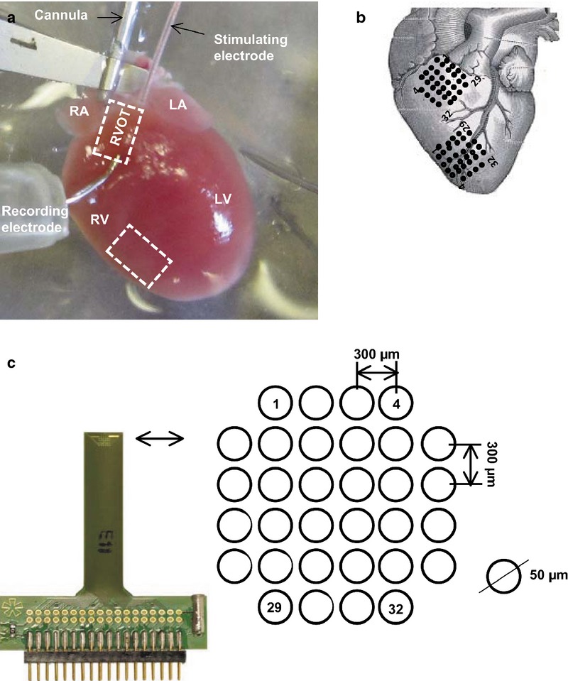
Figure 1.

Electrophysiological experimental set-up for multi-array recordings from the right ventricular outflow tract and right ventricle. (a). Langendorff-perfusion set-up. (b) Placing positions and recording orientations on the free walls of the RVOT and RV for the multi-electrode array (MEA). (c) MEA configuration consisting of 32 electrodes in a square 1.5 × 1.5 mm matrix, with an electrode diameter 50 μm, inter-electrode distance 300 μm. LA, left atrium; RA, right atrium; LV, left ventricle; RV, right ventricle; RVOT, right ventricular outflow tract.
