
FIGURE 2.
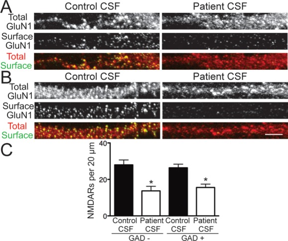
Patient antibodies decreased surface N-methyl-D-aspartate receptors (NMDARs) on excitatory and inhibitory hippocampal neurons. (A, B) Hippocampal neurons immunostained for surface GluN1, total GluN1, and glutamic acid decarboxylase 6 (GAD6) after treatment for 24 hours with control or patient cerebrospinal fluid (CSF) and imaged with confocal microscopy. (A) GAD−, excitatory neurons; (B) GAD+, inhibitory neurons. Surface NMDARs were defined as the colocalization of nonpermeabilized patient antibody staining, which recognized an extracellular epitope, and permeabilized commercial GluN1 staining, which recognized an intracellular epitope. Scale bar = 5μm. (C) Quantification of surface NMDAR density on excitatory, GAD− neurons, and inhibitory, GAD+ neurons (n = 12–28 cells per condition, 3 independent experiments). Treatment with patient CSF caused a similar, significant reduction in surface NMDAR clusters on both excitatory and inhibitory neurons compared to control CSF treatment (excitatory, 27.97 ± 2.67 vs 13.71 ± 2.51; inhibitory, 26.38 ± 1.96 vs 15.6 ± 1.83). *p < 0.05, 1-way analysis of variance.