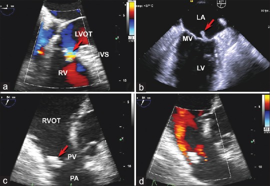
Figure 1.

Transesophageal echocardiography was significant for a very small membranous interventricular septal defect shown in panel (a) with color Doppler demonstrating flow through the ventricular septal defect from the left ventricle to the right ventricle during systole (arrow). Panel (b) demonstrates the mobile vegetation of the posterior leaflet of the mitral valve (arrow). panel (c) shows the large pulmonary valve vegetation (arrow) while panel (d) is a color doppler evaluation of the pulmonary valve demonstrating the pulmonary regurgitation jet in red. IVS = Interventricular septum, LA = Left atrium, LV = Left ventricle, LVOT = Left ventricular outflow tract, PA = Pulmonary artery, PV = Pulmonary valve, RV = Right ventricle, RVOT = Right ventricular outflow tract
