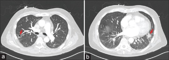
Figure 2.

Chest computed tomography showed evidence of pulmonary infarcts secondary to septic emboli of the pulmonary valve infective endocarditis. Panel (a) shows a nodule in the right upper lobe posterolaterally which is partially cavitated (arrow). panel (b) demonstrates an irregular 17 mm nodule in the lingula posteriorly (arrow)
