


Figure 2.
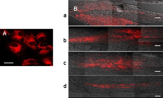
Confocal photomicrographs of PKH26-labeled (red fluorescence) human umbilical cord blood-derived mesenchymal stem cells (hUCB-MSCs) before and after transplantation.
(A) PKH26-labeled hUCB-MSCs before transplantation, captured 2 hours after seeding on a glass slide. Bar: 20 μm.
(B) PKH26-labeled hUCB-MSCs in the lesion site at 1 week (a), 2 weeks (b), 3 weeks (c), and 4 weeks after transplantation (d). Bars: 200 μm.