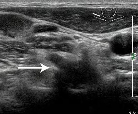
Figure 3.

High-frequency color Doppler ultrasound image of the region where the brachial plexus root traverses the intervertebral foramen in healthy adults.
Nerve root shows an elliptic hypo-echo (arrow). Bony structure of transverse process of vertebra at the region where brachial plexus root traverses the intervertebral foramen is clearly displayed, with strong echo at the bilateral sides of the low-echo structure.
