Abstract
Chagas disease is maintained in nature through the interchange of three cycles: the wild, peridomestic and domestic cycles. The wild cycle, which is enzootic, has existed for millions of years maintained between triatomines and wild mammals. Human infection was only detected in mummies from 4,000-9,000 years ago, before the discovery of the disease by Carlos Chagas in 1909. With the beginning of deforestation in the Americas, two-three centuries ago for the expansion of agriculture and livestock rearing, wild mammals, which had been the food source for triatomines, were removed and new food sources started to appear in peridomestic areas: chicken coops, corrals and pigsties. Some accidental human cases could also have occurred prior to the triatomines in peridomestic areas. Thus, triatomines progressively penetrated households and formed the domestic cycle of Chagas disease. A new epidemiological, economic and social problem has been created through the globalisation of Chagas disease, due to legal and illegal migration of individuals infected by Trypanosoma cruzi or presenting Chagas disease in its varied clinical forms, from endemic countries in Latin America to non-endemic countries in North America, Europe, Asia and Oceania, particularly to the United States of America and Spain. The main objective of the present paper was to present a general view of the interchanges between the wild, peridomestic and domestic cycles of the disease, the development of T. cruzi among triatomine, their domiciliation and control initiatives, the characteristics of the disease in countries in the Americas and the problem of migration to non-endemic countries.
Keywords: ecoepidemiology, Chagas disease, T. cruzi, triatomines domiciliation, control initiatives, endemic and non-endemic countries
The wild cycle of Chagas disease has existed in nature for millions of years. Some accidental human cases of the disease could have occurred, as they still do today, when humans invade the wild ecotope or when wild vectors invade human homes; however human infection by Trypanosoma cruzi has only been identified in mummies of 4,000-9,000 years ago (Guhl et al. 1999, Aufderheide et al. 2004).
Since Carlos Chagas discovered American trypanosomiasis in 1909, the disease that later on received his name, he first observed the domestic and peridomestic cycles in the discovery phase and then the wild cycle of the disease, thereby inferring in a pioneering manner what we know today, that the domestic cycle is a consequence of the wild and peridomestic cycles, respectively.
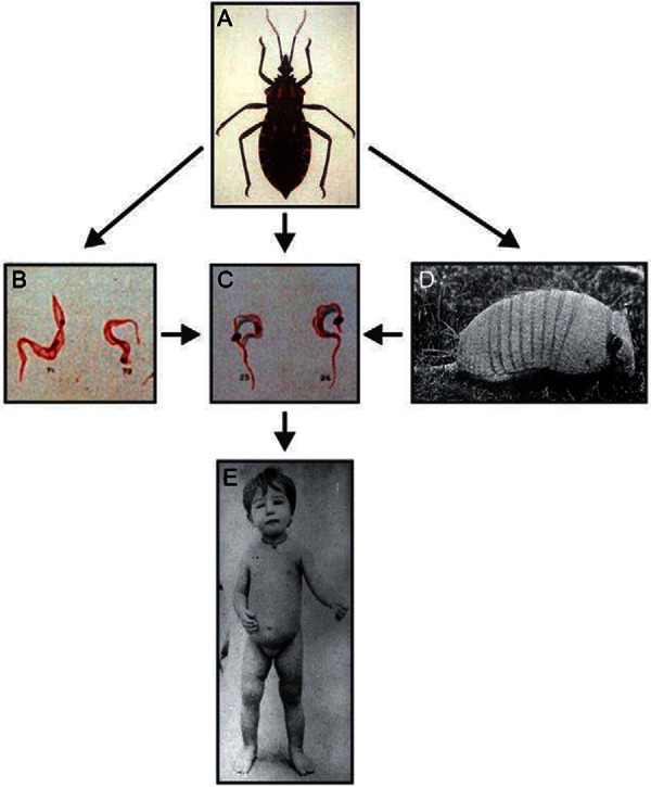
In 1907, Oswaldo Cruz, the director of the Mangui- nhos Institute, where Chagas worked as an assistant researcher, assigned him to work on malaria control, given that this disease was preventing advances in the construction of the Central Railway of Brazil, in northern state of Minas Gerais. Chagas was posted to Lassance, where he occupied a train carriage as his home and laboratory, to start his work on malaria control. In mid-1908, the engineer Cantarino Motta notified Chagas about the presence of an insect called “chupão” (kissing bug) or “barbeiro” (barber) by local inhabitants, which would suck the blood of the residents during the night and hide during the day between cracks in the mud huts where these people lived. By examining the intestinal content of these insects under a microscope, which at that time were called Conorrimus megistus (today known as Panstrongylus megistus), Chagas found “chritidias” (today known as epimastigotes). He deduced that the insects that carried these parasites could transmit them to humans by sucking their blood. Chagas did not have the necessary setup in Lassance to perform experimental studies on animals, so he sent specimens of the infected insect to his director Oswaldo Cruz in Rio de Janeiro, asking him to place them in contact with uninfected marmosets (Callithrix penicillata). Three weeks after placing the insects in contact with the animals, Cruz observed the presence of the parasite in the blood of the marmosets and summoned Carlos Chagas immediately to proceed with the studies on animals. Starting in November 1908, Chagas experimented on mice, guinea pigs, rabbits, dogs and monkeys and confirmed the infection in these mammals. In April 1909, Chagas returned to Lassance, certain he had discovered a new disease. He began examining the people and domestic animals that inhabited the huts in Lassance and neighbouring areas. He first found an infected cat and, on 14 April 1909, he found a child (Berenice), a two-year-old white girl, with fever, hepatosplenomegaly and probably likely an entry-point sign in her eye (later called Romaña’s sign). By examining the child’s blood under a microscope, Chagas found the trypanosome, which he named Schizotrypanum cruzi as a tribute to his master. Thus was discovered American trypanosomiasis. Oswaldo Cruz presented Carlos Chagas’ discovery on 15 April of that year at the National Medical Academy and it was then published in the Memoirs of the Instituto Oswaldo Cruz (Chagas 1909, 1911). In 1912, Carlos Chagas found an armadillo (Dasypus novemcinctus) infected by T. cruzi living in the same burrow as Panstrongylus geniculatus (at that time called Triatoma geniculata). Thus was discovered the wild cycle of Chagas disease (Fig. 1A-E illustrates the discovery of the disease in its domestic and wild cycles). In 1916, Chagas broadened his studies on the acute phase of the disease and, in 1922, Chagas and Villela described the chronic heart form, thereby completing their clinical studies on the disease. Finally, in 1924 Chagas confirmed T. cruzi as the parasite found in naturally infected Chrisotrix ciureus (today known as Saimiri sciureus) monkeys in the state of Pará, thus consolidating the knowledge of the wild cycle of the disease.
Fig. 1. : the discovery of Chagas disease (original from Carlos Chagas). A: Panstrongylus megistus (Chagas 1909); B, C: Trypanosoma cruzi (Chagas 1909); D: armadillo Dasypus novemcinctus (Chagas 1912); E: acute cases of Chagas disease (Chagas 1916).

The developmental process of T. cruzi in triatomines and their domiciliation - More than 140 species of triatomines have been recognised, grouped into 19 genera and five tribes. Among these, the vast majority are wild and associated with the ecotopes of mammals and birds; some are within peridomestic areas, such as in chicken coops, pigsties and corrals and a few are found within homes, thus constituting important vectors for humans. Among this last group is Triatoma infestans in the southern cone of South America and Rhodnius prolixus in the Andean Region and in Central America (Galvão et al. 2003, Catalá et al. 2004, Villela et al. 2005). Although triatomines have been known since the XVI century (Lent & Wigodzinski 1979), they were only formally described by De Geer in 1773 and infection by T. cruzi was only first described by Carlos Chagas in 1909. According to Hoare (1972), Léger was the first to speculate on the phylogenetic relationships among trypanosomes, suggesting, in 1904, that ancestral trypanosomes were monogenetic insects that did not suck blood. When triatomines began sucking the blood of mammals, trypanosomes underwent morphological modifications, such as flagellum and undulating membrane, in order to circulate in the mammal’s blood. This hypothesis has been adopted and broadened in its essence by several authors (Hoare 1972). The evolution of T. cruzi according to the “bat seeding hypothesis” the bat trypanosomes have switched into terrestrial mammals of South America (Hamilton et al. 2012).
The process of domiciliation or domestication of the triatomines is still not entirely known. The evolution of haematophagy in the Triatominae family is believed to have evolved from several lineages of Reduviidae, which firstly becoming adapted as optional blood-sucking predators, followed by obligate haematophagy in vertebrate niches (Schofield 2000). The evolution from predators to mandatory blood-suckers was associated with a series of morphological modifications related with three key factors: (i) the use of a vertebrate’s blood as a food source, (ii) adaptation to the host’s environment and (iii) a progressive dependency on the host as a means of dispersion through passive carriage. Two factors are fundamental for demographic stability among triatomines: (i) stability of the food source and (ii) protection from climatic extremes. On the other hand, passive carrying of triatomines by vertebrates explains how the domestic species, such as T. infestans and R. prolixus, have become disseminated. Secondary factors for dispersion include the ability to fly among adult triatomines and attraction to ultraviolet light (Schofield et al. 1999).
Finally, the preponderant factor for peridomiciliation and domiciliation of triatomines is uncontrolled deforestation with the removal of natural food sources, especially wild mammals, which compels insects to come closer to human peridomestic and domestic areas in search of the food sources that became lost through banishment of wild mammals and birds. This is the major risk domiciliation of triatomines in the Amazon Region (Coura & Junqueira 2012).
Several phenotypic and genetic studies on different species in different ecotopes have demonstrated variations among these species, regarding the sensilla of the insects’ antenna, the size of the head and wings, genetic simplification in relation to wild, peridomestic and domestic habits and geographical distribution (Dujardin 1998, Catalá & Dujardin 2001, Cabajal de la Fuente & Catalá 2002, Abraham et al. 2008, Hernández et al. 2008, 2011, 2011, 2013, Cabajal de la Fuente et al. 2009, Gorla et al. 2013).
Epidemiological characteristics of Chagas disease in the Americas - Chagas disease in the Americas can be separated into four groups according to the wild, peridomestic and domestic cycles and the situation of human infection and disease (Coura & Dias 2009, Coura et al. 2009).
Group I - This group includes Argentina, Bolivia, Brazil, Chile, Ecuador, Paraguay, Peru and Venezuela, which are characterised by the wild, peridomestic and domestic cycles with high prevalence zones for human infection predominance of heart disease, with an absence of or rare cases of the digestive form northwards from the equator. The wild cycle is frequently found in several environments within this group and other groups of countries, including T. infestans in Bolivia and Peru. Control over the transmission of infection by T. infestans was certified by the Pan-American Health Organization (PAHO) in Uruguay in 1997, in Chile in 1999 and in Brazil in 2006. In Brazil still remain two isolated foci of T. infestans: one in the state of Rio Grande do Sul and the other in the state of Bahia. These countries have also controlled T. cruzi transmission through blood transfusions. Argentina and Paraguay are still working towards this goal, but so far have not been able to eliminate T. infestans as a main vector, particularly in the Gran Chaco region (Gorla et al. 2013).
Group II - Group II includes Colombia, Costa Rica and Mexico, which are characterised by the peridomestic and domestic cycles, with the presence of chronic Chagas heart disease, occurrences of blood donors who have been infected by T. cruzi and an incipient vector control, naturally with the wild cycle in its own environment.
Group III - This group includes El Salvador, Guatemala, Honduras, Nicaragua and Panama with the presence of the wild, peridomestic and domestic cycles, with very limited information on the clinical forms of the disease. However, vector control actions are underway in El Salvador, Guatemala, Honduras and Nicaragua (Hashimoto & Schofield 2012).
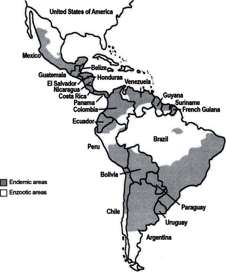
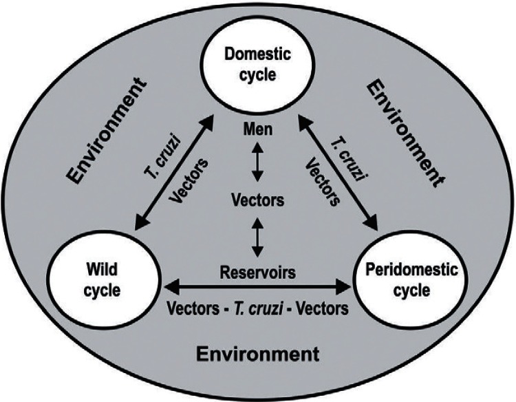
Group IV - This group includes the Antilles, the Bahamas, Belize, Cuba, the United States of America (USA), Guyana, French Guiana, Haiti, Jamaica and Surinam, where the wild cycle is predominant, with several cases found among immigrants from endemic countries who live in the USA (Schmunis 2007, Coura & Albajar-Viñas 2010), where control over blood donors is incipient and is being broadened. There have been reports of cases cause by blood transfusion (Zeledón et al. 2012). Fig. 2 shows the endemic and enzootic areas of Chagas disease in the Americas (WHO 1991) and Fig. 3 illustrates the interchange of the wild, peridomestic and domestic cycles (Coura & Dias 2009).
Fig. 2. : endemic and enzootic areas of Chagas disease in the Americas. Up to date: Triatoma infestans was eliminated from Uruguay, Chile and Brazil and is under control in Argentina and Paraguay, and Rhodnius prolixus is under control in Central America.
Fig. 3. : interchange of the wild, peridomestic and domestic cycles (Coura & Dias 2009).
Initiative for controlling Chagas disease in the Americas - Multinational initiatives for controlling Chagas disease have been created in the Southern Cone, the Andean countries, Central America and Mexico and in the Amazon Region since 1991.
Southern Cone Initiative (INCOSUR) - The ministries of health of Argentina, Bolivia, Brazil, Chile, Paraguay and Uruguay met in Brasília in July 1991 to participate in a Regional Assembly of Health, including discuss strategies for controlling Chagas disease. Thus, they created an “intergovernmental commission” for controlling the disease, with the PAHO forming the secretariat, with a subregional design and an action plan to eliminate T. infestans and interrupt the transmission of T. cruzi through blood transfusion. This effort particularly needed to take into account for the context of each country, the existence of national plans and technical cooperation between the countries (Coura et al. 2009). The goals of the subregional INCOSUR were: (i) elimination of T. infestans from the domestic and peridomestic areas of endemic regions, (ii) reduction or elimination of other species of domestic triatomines in the same areas that were endemic for T. infestans and (iii) elimination of T. cruzi transmission through blood transfusion, by means of screening donors at blood banks. The elimination of T. infestans was certified by the PAHO in Uruguay in 1997, in Chile in 1999 and in Brazil in 2006, as was elimination of T. cruzi transmission through blood transfusion in these countries. Argentina and Paraguay have controlled T. infestans in some provinces, while Bolivia has reduced vector transmission very little, particularly in the Gran Chaco region, which is divided between these three countries, Argentina, Bolivia and Paraguay (Coura 2013). Recently, Abad-Franch et al. (2013) discussed the certification of the interruption of Chagas disease transmission by native vectors.
Initiative of the Andean Countries (IAC) - The IAC was officially launched by Colombia, Ecuador, Peru and Venezuela in 1997 (Guhl 2007). The high ecological diversity, the diversity of triatomine species and the great complexity of Chagas disease pose a great challenge controlling in this area (Coura et al. 2009). R. prolixus is one of the main domestic vectors in Venezuela, where a wild population continues to exist in palm trees, in some of the other Andean states, where can be confused with Rhodnius robustus, but it re-infests human homes, thus creating a challenge with regard to controlling Chagas disease. Triatoma maculata is a secondary domestic vector in some areas of Venezuela. In Colombia, the main goal of the vector control programme is to interrupt transmission by R. prolixus, which is the main domestic vector in that country. Triatoma dimidiata and, to a lesser extent, Rhodnius pallescens are potential vectors as well. In Ecuador, the goal is to eliminate T. dimidiata, which is a vector that was artificially introduced into the country, as well as Rhodnius ecuadoriensis on the west coast and in the Andean valleys. This vector is also present in homes in northern Peru, but this population is genetically different from those of Ecuador (Guhl 2007). Also in Peru, Panstrongilus herreri is an important vector in the valley of the Marañon River. Finally, in southern Peru, elimination of the population of T. infestans is the maximum priority.
Initiative of Central America and Mexico - The Central America Initiative was created based on a resolution adopted during a meeting in Belize, in September 1997, which was consolidated in October of that year in Tegucigalpa, Honduras, where the following objectives were established: (i) to interrupt the transmission of Chagas disease by R. prolixus, which needed to be eliminated from the region, (ii) to reduce domestic infestation by T. dimidiata and (iii) to interrupt T. cruzi transmission through blood transfusion (Coura et al. 2009).
In Guatemala, 100% of the homes within the area infested by R. prolixus were sprayed with insecticides, with the aid of the Japanese International Cooperation Agency, thus interrupting the transmission via this vector in 2008. In El Salvador, housing improvements, associated with controlling the malaria vector by means of insecticides, resulted in elimination of R. prolixus and consequently elimination of T. cruzi transmission by the vector, which was no longer found in that country. About 90% of the territory of Honduras is considered to be subject to the presence of triatomines. However, R. prolixus has only been found in very poor homes. T. dimidiata, which is native to Central America, can be wild, peridomestic or domestic and despite being considered to be a secondary vector, it is certainly the biggest problem regarding Chagas disease control in this region in the near future. The control programme for R. prolixus in Honduras foresaw the elimination of this vector in 2010. According to a recent evaluation, El Salvador, Guatemala, Honduras and Nicaragua can be considered to have been successful in eliminating R. prolixus, although isolated foci of this species may still exist. Moreover, the evaluation showed that in Belize, Costa Rica and Panama, this vector no longer exists. In Panama, homes are instead invaded by R. pallescens, which infests the palm trees in peridomestic areas. Given that there have been positive serological findings in children, an extensive entomological survey covering all the countries of Central America becomes necessary in order to make a more evaluation of the current situation of Chagas disease in the region. Mexico is certainly a country that needs strong encouragement to begin a more effective programme for Chagas disease control and such initiatives have recently been aroused (Zeledón et al. 2006, Ponce 2007, Guhl 2007, Coura et al. 2009, Coura 2013). Hashimoto and Schofield (2012) discussed the elimination of R. prolixus from Central America and Carabarin-Lima et al. (2013) have presented an update on Chagas disease in Mexico.
Initiative of the Amazon Countries (AMCHA) - In a preliminary meeting held in Palmarí, in the municipality of Tabatinga on the Solimões River (Brazil), on the border with Letícia (Colombia), between 30 July-2 August 2002, the problems of Chagas disease were discussed by a group of international scientists convoked by the European Community and Latin America Triatominae Network, the need to create an intergovernmental initiative among the nine Amazon countries in order to monitor and prevent the disease in those countries. The participants in the meeting in Palmarí suggested that a new meeting should be held to create the AMCHA, which would be organised by the PAHO. This meeting occurred in Manaus (Brazil), 19-22 September 2004, and had the following objectives: (i) to evaluate the risks of Chagas endemicity becoming established in the Amazon Region, (ii) to identify the research required for monitoring and prevention of Chagas disease in the Amazon Region, (iii) to propose monitoring and prevention for Chagas disease in this region and (iv) to propose of an international cooperation system for Chagas disease monitoring and prevention in the Amazon Region (AMCHA). At this meeting promoted by the PAHO, the nine Amazon countries were represented: Bolivia, Brazil, Colombia, Ecuador, Guyana, French Guiana, Peru, Surinam and Venezuela. In addition to the official representatives from these different countries, more than 90 researchers from several Amazon countries participated in the meeting and discussed in detail the problems of Chagas disease in the region, the objectives of AMCHA and how to put AMCHA into operation.
Three other meetings were held, respectively in Cayenne, French Guiana, 2-4 November 2005, in Quito, Ecuador, 18-20 September 2006, and in Caracas, Venezuela, 28-30 October 2008. However, despite recognition of the risks of endemic Chagas disease in the Amazon Region, the need to implement research in the region and to gain deeper knowledge of this endemic disease and the various proposals for monitoring and prevention of the disease, very little concrete action has been taken in spite of the great efforts made by the PAHO. It was expected that through the meeting in Bolivia, in 2012, and the next meeting scheduled for August 2014 in the state of Acre, Brazil, there would be an increase in international cooperation for surveillance and prevention of Chagas disease in the Pan-Amazon region and that the AMCHA initiative would effectively fulfil the goals that had been proposed at its founding meeting in Manaus, Brazil, in September 2004.
Most acute cases of Chagas disease in the Brazilian Amazon Region are caused by outbreaks of oral transmission (Valente et al. 1999, 2009, Pinto et al. 2008, Souza-Lima et al. 2013) and vector transmission is almost always attributed to plant extraction activities, particularly those involving piassava. High frequencies of chronic cases of the disease (Coura et al. 2002, Albajar-Viñas et al. 2003, Xavier et al. 2006, Brum-Soares et al. 2010) or accidental cases can occur in places where humans penetrates the forests for a variety of reasons and also where vectors and wild animals (marsupials) invade human homes in search of food. Thus, the perspectives on the prevention and control of Chagas disease in this region are necessarily different from the techniques applied to the classical endemic areas, in which the vectors have become domesticated and transmission occurs continuously. In the Amazon Region, where there is no vector adaptation (with the exceptions mentioned earlier), it is fundamentally important to train technicians to be able to deliver early diagnosis of acute cases, to apply treatment and to recognise triatomines during epidemiologic surveillance. Information that is provided intensively to the population through posters, radio, television, health agents and elementary school teachers and by means of courses and seminars to train surveillance multipliers, is indispensable. Moreover, training laboratory technicians to make early diagnoses of acute cases of the disease and to recognise triatomines is paramount for epidemiological surveillance, as is training doctors and nurses to make clinical diagnoses, to provide treatment for acute and chronic cases of Chagas disease and to implement disease control in the Amazon Region.
The experience of our group, with capacitation courses for malaria microscopists and laboratory technicians for the public health network, so that they are able to detect T. cruzi in routine tests for diagnosing malaria and to carry out blood cell counts and direct T. cruzi investigations through fresh blood examination or thick blood smears in cases in which Chagas infection is suspected and also to recognise triatomines and examine their faeces so as to detect T. cruzi have been shown to be of great importance for the surveillance of Chagas disease in the Brazilian Amazon Region. Courses held in the nine Brazilian states in the Amazon Region, with the support of Médecins Sans Frontières between 2006-2008, were of prime importance for detecting acute cases of the disease in the region, with an annual notification of 100-150 cases per year ever since the courses began. Naturally, this is only a small proportion of the occurrences, since most individuals with the disease do not seek the public health and hospital services of the region. These occurrences are certainly 10-20 times more frequent, taking undetected cases into consideration. On the other hand, several acute outbreaks of the disease have been identified in the region, partly thanks to the dissemination about the disease. As a consequence of these courses, a Technician Capacitation Manual for the detection of T. cruzi was created with financial aid from the PAHO, using resources from the WHO Chagas Disease Programme, and this manual was then improved for a second edition CDs that was widely distributed in the Brazilian Amazon Region. An edition in Spanish is also being prepared for distribution in Spanish-speaking countries.
The courses and the respective manual were elaborated by ACV Junqueira, TCM Gonçalves and CJ Carvalho-Moreira revised by JR Coura and several collaborators, in a corrected second edition (2011), financed through the project Ecoepidemiology of Chagas Disease in the Amazon Region, by Department of Science and Technology (Ministry of Health) and Council for Scientific and Technological Development, with backing from the PAHO/WHO Neglected Infectious Diseases Programme and coordination by the Parasitic Diseases Laboratory, Oswaldo Cruz Institute, Oswaldo Cruz Foundation (Coura et al. 2011).
Chagas disease in non-endemic countries - A new epidemiological, economic and social problems have been created through the globalisation of Chagas disease, due to the legal and illegal migration of individuals infected by T. cruzi or presenting Chagas disease in its varied clinical forms, from endemic countries in Latin America to non-endemic countries in North America, Europe, Asia and Oceania, and particularly to the USA, Canada, Spain, Switzerland, Japan and emerging countries in Asia, Australia and other countries in Oceania (Schmunis 2007, Coura & Dias 2009, Coura et al. 2009, Coura & Albajar 2010). These migrants have created a new epidemiological and public health problem for the countries that received them. These problems include the risks of infection transmission through blood transfusion or congenitally and the need for greater control over blood donors and medical care for patients in countries with little experience with this subject. On the other hand, there are also economic and social issues relating to migration control, patient treatment, epidemiological surveillance and reduction of the manpower needed in more developed countries.
An estimated 300,000 legal immigrants from Latin America in the USA are infected by T. cruzi (Bern & Montgomery 2009, Coura & Albajar Viñas 2010). However, in the southern USA, and especially in Texas, the wild cycle of the disease was established a long time ago: some autochthonous acute cases of Chagas disease occur occasionally and human homes are invaded by wild triatomines frequently (Zeledón et al. 2012). From 1955-2012, only 23 autochthonous cases of Chagas disease were reported in the USA (Wood & Wood 1955, Cantey et al. 2012, Montgomery et al. 2014). Serological screening studies carried out between January 2007-January 2013 in the USA identified 1,752 blood donors who were positive for T. cruzi infection, among whom more than 100 were from Texas. An estimated 63-315 children have acquired T. cruzi infection congenitally in the USA (Bern e Montgomery 2009, Montgomery et al. 2014). Similarly, perinatal screening on 4,000 women, mostly of Hispanic origin, revealed that 0.25% were infected by T. cruzi and thus presented a risk of congenital transmission of the disease (Edwards et al. 2013). On the other hand, Bern and Montgomery (2009) and Montgomery et al. (2014) estimated that approximately 30,000-45,000 people in the USA have Chagas cardiomyopathy.
There are an estimated three million migrants from Latin America in the following European countries: Belgium, France, Germany, Italy, the Netherlands, Portugal, Spain and Switzerland. Among these migrants, there are an estimated 68,000-123,000 cases of Chagas disease. However, only a small proportion of these individuals have been diagnosed. Up to 2009, only 4,300 cases had been diagnosed, of which 89% were in Spain (Basile et al. 2011).
Until the beginning of the 1980s, few cases of Chagas disease transmitted by blood transfusion, transmitted congenitally or acquired in a laboratory, among immigrant workers, had been published in Western Europe (Albajar-Viñas & Jannin 2011). In the mid-1990s, there was a marked increase in the number of immigrants from Latin America to Europe, especially to Spain, which consequently led to an increased number of cases, which were mostly asymptomatic in the chronic phase of the disease. This situation led to changes to the legislation regarding screening for blood donors for transfusion and tissue donors for transplantation, due to the risks of transmitting the disease. A new estimate has established that Spain will still have 29,000 cases of chronic Chagas heart disease in the near future (Perez de Ayala 2009).
Footnotes
Financial support: FIOCRUZ, CNPq
REFERENCES
- Abad-Franch F, Diotaiuti L, Gurgel-Gonçalves R, Gürtler RE. Certifying the interruption of Chagas disease transmission by native vectors: cui bono? Mem Inst Oswaldo Cruz. 2013;108:251–254. doi: 10.1590/0074-0276108022013022. [DOI] [PMC free article] [PubMed] [Google Scholar]
- Abraham L, Hernandez L, Gorla D, Catalá S. Phenotypic of Triatoma infestans in the microgeographic level in the Gran Chaco of Argentina and the Andean valleys of Bolivia. J Med Entomol. 2008;45:660–666. doi: 10.1603/0022-2585(2008)45[660:pdotia]2.0.co;2. [DOI] [PubMed] [Google Scholar]
- Albajar-Viñas P, Jannin J. The hidden Chagas disease in Europe. Euro Surveill. 2011;16:1–2. doi: 10.2807/ese.16.38.19975-en. [DOI] [PubMed] [Google Scholar]
- Albajar-Viñas P, Loredo SV, Terrazas MB, Coura JR. Miocardiopatia dilatada em pacientes com infecção chagásica crônica. Relato de dois casos fatais autóctones do Rio Negro, estado do Amazonas. Rev Soc Bras Med Trop. 2003;36:401–407. [PubMed] [Google Scholar]
- Aufderheide AC, Salo W, Maden M, Streitz J, Bulkestra J, Guhl F, Amaza B, Renier C, Wittmers LE, Jr, Formaciari G, Alison A. A 9,000-years record of Chagas disease. Proc Nat Acad Sci USA. 2004;101:2034–2039. doi: 10.1073/pnas.0307312101. [DOI] [PMC free article] [PubMed] [Google Scholar]
- Basile L, Jansen J, Carlier Y, Salamanca D, Amgheben A, Bartoloni A. Chagas disease in European countries: the challenge of a surveillance system. Euro Surveil. 2011;16 [PubMed] [Google Scholar]
- Bern C, Montgomery SP. An estimate of the burden of Chagas disease in the United States. Clin Infect Dis. 2009;49:52–54. doi: 10.1086/605091. [DOI] [PubMed] [Google Scholar]
- Brum-Soares LM, Xavier SS, Sousa AS, Pereira JB, Junqueira ACV, Coura JR. Morbidade da doença de Chagas em pacientes autóctones da microrregião do Rio Negro, estado do Amazonas. Rev Soc Bras Med Trop. 2010;43:170–177. doi: 10.1590/s0037-86822010000200013. [DOI] [PubMed] [Google Scholar]
- Cabajal de la Fuente AL, Catalá S. Relationship between antennal sensilla pattern and habitat in six species of Triatominae. Mem Inst Oswaldo Cruz. 2002;97:1121–1125. doi: 10.1590/s0074-02762002000800010. [DOI] [PubMed] [Google Scholar]
- Cabajal de la Fuente AL, Porcasi X, Noireau F, Diotaiuti L, Gorla DE. The association between the geographic distribution of Triatoma pseudomaculata and Triatoma wygodzinskyi (Hemiptera: Reduviidae) with environment variables recorded by remote sensors. Infect Genet Evolut. 2009;9:54–61. doi: 10.1016/j.meegid.2008.09.008. [DOI] [PubMed] [Google Scholar]
- Cantey PT, Stramer SL, Towsend RL, Kamel H, Ofafa K, Todd PL, Montgomert SP. The United State Trypanosoma cruzi Infection Study: evidence for vector borne transmission of the parasite that causes Chagas disease among United States blood donors. Transfusion. 2012;52:1922–1930. doi: 10.1111/j.1537-2995.2012.03581.x. [DOI] [PubMed] [Google Scholar]
- Carabarin-Lima A, González-Vasquez MC, Rodriguez-Moralez O, Baylon-Pacheco L, Rozales-Encina JL, Reyes-López PA, Arce-Fonseca M. Chagas disease (American trypanosomiasis) in Mexico: an update. Acta Tropica. 2013;127:126–135. doi: 10.1016/j.actatropica.2013.04.007. [DOI] [PubMed] [Google Scholar]
- Catalá S, Dujardin JP. Antennal sensilla patterns indicate geographic and ecotopic variability among Triatoma infestans (Hemiptera: Reduviidae) populations. J Med Entomol. 2001;38:423–428. doi: 10.1093/jmedent/38.3.423. [DOI] [PubMed] [Google Scholar]
- Catalá SS, Maida DM, Caro-Riaño H, Jaramillo N, Moreno J. Changes associated with laboratory rearing in antennal sensilla patterns of Triatoma infestans, Rhodnius prolixus and Rhodnius pallescens (Hemiptera, Reduviidae, Triatominae) Mem Inst Oswaldo Cruz. 2004;99:25–30. doi: 10.1590/s0074-02762004000100005. [DOI] [PubMed] [Google Scholar]
- Chagas C. Nova tripanozomiaze humana. Estudos sobre a morfolojia e o ciclo evolutivo do Schizotrypanum cruzi n. gen., n. sp., ajente etiolojico de nova entidade morbida do homem. Mem Inst Oswaldo Cruz. 1909;1:159–218. [Google Scholar]
- Chagas C. Nova entidade morbida do homem. Rezumo geral de estudos etiolojicos e clinicos. Mem Inst Oswaldo Cruz. 1911;3:219–275. [Google Scholar]
- Chagas C. Sobre um Trypanosoma do tatu, Tatusia novemcineta, transmitido pelo Triatoma geniculata Latr (1811). Possibilidade de ser o tatu um depositário do Trypanosoma cruzi no mundo exterior. Nota prévia. Braz Med. 1912;26:305–336. [Google Scholar]
- Chagas C. Tripanosomiase americana. Forma aguda da moléstia. Mem Inst Oswaldo Cruz. 1916;8:37–65. [Google Scholar]
- Chagas C. Infection naturelle dês singe du Pará (Chrysotrix sciureus) par Trypanosoma cruzi. Comp Rend Séanc Soc Biol Ses Fin. 1924;90:873–876. (Fre). [Google Scholar]
- Chagas C, Villela E. Forma cardíaca da tripanosomíase americana. Mem Inst Oswaldo Cruz. 1922;14:5–61. [Google Scholar]
- Coura JR. Chagas disease: control, elimination and eradication. Is it possible? Mem Inst Oswaldo Cruz. 2013;108:962–967. doi: 10.1590/0074-0276130565. [DOI] [PMC free article] [PubMed] [Google Scholar]
- Coura JR, Abad-Franch F, Aguilera X, Dias JCP, Gil E, Junqueira ACV, Fiusa-Lima J, Carvalho-Moreira CJ, Schall VT, Schmunis G. The initiatives for the control of Chagas disease in the Americas and in non-endemic countries: overview and perspectives. Rev Soc Bras Med Trop. 2009;42(Suppl II):106–110. [Google Scholar]
- Coura JR, Albajar-Viñas P. Chagas disease: a new worldwide challenge. Nature. 2010;465(Suppl):S6–S7. doi: 10.1038/nature09221. [DOI] [PubMed] [Google Scholar]
- Coura JR, Dias JCP. Epidemiology, control and surveillance of Chagas disease - 100 years after its discovery. Mem Inst Oswaldo Cruz. 2009;104(Suppl. I):31–40. doi: 10.1590/s0074-02762009000900006. [DOI] [PubMed] [Google Scholar]
- Coura JR, Junqueira ACV. Risks of endemicity, morbidity and perspectives of regarding the control of Chagas disease in the Amazon Region. Mem Inst Oswaldo Cruz. 2012;107:145–154. doi: 10.1590/s0074-02762012000200001. [DOI] [PubMed] [Google Scholar]
- Coura JR, Junqueira ACV, Fernandes O, Valente SAS, Miles MA. Emerging Chagas disease in Amazonian Brazil. Trends Parasitol. 2002;18:171–176. doi: 10.1016/s1471-4922(01)02200-0. [DOI] [PubMed] [Google Scholar]
- Coura JR, Junqueira ACV, Gonçalves TCM, Carvalho Moreira CJ. Manual de capacitação na detecção de Trypanosoma cruzi para microscopistas de malária e laboratoristas de rede de saúde pública. OPAS/OMS-Fiocruz; Rio de Janeiro: 2011. 284 [Google Scholar]
- Dujardin JP. Population genetics and the natural history of domestication in Triatominae. Mem Inst Oswaldo Cruz. 1998;93(Suppl. II):34–36. [Google Scholar]
- Edwards MS, Rench MA, Todd CW, Czaicki N, Steurer FJ, Bern C, Montgomery SP. Perinatal screening for Chagas disease in southern Texas. 10.1093/ipids/pit056J Ped Infect Dis Soc. 2013 doi: 10.1093/jpids/pit056. [DOI] [PMC free article] [PubMed] [Google Scholar]
- Galvão C, Carvalho RU, Rocha DS, Juberg J. A check-list of the current valid species of the sub-family Triatominae Jeannel, 1919 (Hemiptera: Reduviidae) and their geographical distribution with nomenclatural and taxonomic notes. Zootaxa 2002. 2003:1–36. [Google Scholar]
- Gorla DE, Abrahan L, Hernández ML, Porcasi X, Hrellac HÁ, Carrizo H, Catalá SS. New structures for goat corrals to control peridomestic populations of Triatoma infestans (Hemiptera: Reduviidae) in the Gran Chaco of Argentina. Mem Inst Oswaldo Cruz. 2013;108:352–358. doi: 10.1590/S0074-02762013000300014. [DOI] [PMC free article] [PubMed] [Google Scholar]
- Guhl F. Chagas disease in Andean countries. Mem Inst Oswaldo Cruz. 2007;102(Suppl. I):29–37. doi: 10.1590/s0074-02762007005000099. [DOI] [PubMed] [Google Scholar]
- Guhl F, Jamillo C, Vallejo GA, Voekteng R, Cardenas-Arroyo F, Forniciari C. Isolation of Trypanosoma cruzi DNA in 4,000 years old mummified human tissue from northern Chile. Am J Physiol Anthropol. 1999;108:625–635. doi: 10.1002/(SICI)1096-8644(199904)108:4<401::AID-AJPA2>3.0.CO;2-P. [DOI] [PubMed] [Google Scholar]
- Hamilton PB, Teixeira MMG, Stevens JR. The evolution of Trypanosoma cruzi: the “bat seeding” hypothesis. Trends Parasitol. 2012;28:136–141. doi: 10.1016/j.pt.2012.01.006. [DOI] [PubMed] [Google Scholar]
- Hashimoto K, Schofield CJ. Elimination of Rhodnius prolixus in Central America. 45Parasit Vectors. 2012;5 doi: 10.1186/1756-3305-5-45. [DOI] [PMC free article] [PubMed] [Google Scholar]
- Hernández L, Abraham B, Moreno M, Gorla D, Catalá S. Phenotypic variability associated to genomic changes in the main vector of Chagas disease in the Southern Cone of South America. Acta Tropica. 2008;106:60–67. doi: 10.1016/j.actatropica.2008.01.006. [DOI] [PubMed] [Google Scholar]
- Hernández ML, Abraham LB, Dujardin JP, Gorla DE, Catalá SS. Phenotypic variability and population structure of peridomestic Triatoma infestans in rural areas of arid Chaco (Western Argentina): spatial influence of macro and microhabitants. Vector Borne Zoonotic Dis. 2011;11:503–513. doi: 10.1089/vbz.2009.0253. [DOI] [PubMed] [Google Scholar]
- Hernández ML, Dujardin JP, Gorla DE, Catalá SS. Potential sources of Triatoma infestans reinfesting peridomiciles identified by morphological characterization in los Llanos, La Rioja, Argentina. Mem Inst Oswaldo Cruz. 2013;108:91–97. doi: 10.1590/S0074-02762013000100015. [DOI] [PMC free article] [PubMed] [Google Scholar]
- Hoare CA. Blackwell Scientific Publication. Oxford-Edinburgh; 1972. The trypanosomes of mammals. A zoological monograph.749 [Google Scholar]
- Lent H, Wigodzinsky P. Revision of triatomines (Hemiptera: Reduviidae) and their significance as vector of Chagas disease. Bull Am Mus Nat History. 1979;63:125–520. [Google Scholar]
- Montgomery SP, Starr MC, Cantey PT, Edwards MS, Meymandi SK. Neglected parasitic infections in the United States. Am J Trop Med Hyg. 2014;90:814–818. doi: 10.4269/ajtmh.13-0726. [DOI] [PMC free article] [PubMed] [Google Scholar]
- Perez de Ayala A, Perez-Moliona JA, Norman F, Lopez-Velez R. Chagasic cardiomyopathy in imigrants from Latin America to Spain. Emerg Infect Dis. 2009;15:607–608. doi: 10.3201/eid1504.080938. [DOI] [PMC free article] [PubMed] [Google Scholar]
- Pinto AYN, Valente SAS, Valente VC, Ferreira AG, Junior, Coura JR. Fase aguda da doença de Chagas na Amazônia brasileira. Estudo de 233 casos no Pará, Amapá e Maranhão observados entre 1988 e 2005. Rev Soc Bras Med Trop. 2008;41:602–614. doi: 10.1590/s0037-86822008000600011. [DOI] [PubMed] [Google Scholar]
- Ponce C. Current situation of Chagas disease in Central America. Mem Inst Oswaldo Cruz. 2007;102(Suppl. I):41–44. doi: 10.1590/s0074-02762007005000082. [DOI] [PubMed] [Google Scholar]
- Schmunis GA. Epidemiology of Chagas disease in non-endemic countries: the role of international migration. Mem Inst Oswaldo Cruz. 2007;102(Suppl. I):75–85. doi: 10.1590/s0074-02762007005000093. [DOI] [PubMed] [Google Scholar]
- Schofield CJ. Trypanosoma cruzi - the vector-parasite paradox. Mem Inst Oswaldo Cruz. 2000;95:535–544. doi: 10.1590/s0074-02762000000400016. [DOI] [PubMed] [Google Scholar]
- Schofield CJ, Diotaiuti L, Dujardin JP. The process of domestication in Triatominae. Mem Inst Oswaldo Cruz. 1999;94(Suppl. I):375–378. doi: 10.1590/s0074-02761999000700073. [DOI] [PubMed] [Google Scholar]
- Souza-Lima RC, Barbosa MGV, Coura JR, Arcanjo ARL, Nascimento AS, Ferreira JMBB, Magalhães IK, Albuquerque BV, Araujo GAN, Guerra JAO. Outbreak of acute Chagas disease in the Rio Negro region. Rev Soc Bras Med Trop. 2013;46:510–514. doi: 10.1590/0037-8682-1367-2013. [DOI] [PubMed] [Google Scholar]
- Valente SAS, Valente VC, Fraiha H., Neto Considerations on the epidemiology and transmission of Chagas disease in the Brazilian Amazon. Mem Inst Oswaldo Cruz. 1999;94(Suppl. I):395–398. doi: 10.1590/s0074-02761999000700077. [DOI] [PubMed] [Google Scholar]
- Valente SAS, Valente VC, Pinto AYN, César MIR, Santos MP, Miranda COS, Cuervo P, Fernandes O. Analysis of an acute Chagas disease outbreak in the Brazilian Amazon: human cases, triatomines, reservoir mammals and parasites. Trans R Soc Trop Med Hyg. 2009;103:291–297. doi: 10.1016/j.trstmh.2008.10.047. [DOI] [PubMed] [Google Scholar]
- Villela MM, Catalá S, Jurberg J, Silva IG, Dias JCP. Patterns of antenal sensilla of Panstrongylus megistus from three Brazilian states. Mem Inst Oswaldo Cruz. 2005;100:699–702. doi: 10.1590/s0074-02762005000700002. [DOI] [PubMed] [Google Scholar]
- WHO - World Health Organization Control of Chagas disease. WHO Tech Rep Ser. 1991;811:1–95. [PubMed] [Google Scholar]
- Woody NC, Woody HP. American trypanosomiasis (Chagas disease): first indigenous case in the United States. JAMA. 1955;159:676–677. doi: 10.1001/jama.1955.02960240042010a. [DOI] [PubMed] [Google Scholar]
- Xavier SS, Souza AS, Albajar PV, Junqueira ACV, Bóia MN, Coura JR. Cardiopatia chagásica crônica no Rio Negro, estado do Amazonas. Relato de três novos casos autoctónes comprovados por exames sorológicos, clínicos, radiográficos de tórax, eletro e ecocardiográficos. Rev Soc Bras Med Trop. 2006;39:211–216. doi: 10.1590/s0037-86822006000200015. [DOI] [PubMed] [Google Scholar]
- Zeledón R, Beard CB, Dias JCP, Leiby DA, Dorn PL, Coura JR. An appraisal of the status of Chagas disease in the United States. Elsevier: Amsterdan/Oxford; 2012. 102 [Google Scholar]
- Zeledón R, Marin F, Calvo N, Lugo E, Vale S. Distribution and ecological aspects of Rhodnius pallescens in Costa Rica and Nicaragua and their epidemiological implication) Mem Inst Oswaldo Cruz. 2006;101:75–79. doi: 10.1590/s0074-02762006000100014. [DOI] [PubMed] [Google Scholar]


