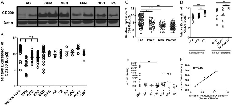
Figure 1.

Brain tumors express CD200. A. Western analysis of CD200 on Anaplastic oligoastrocytoma; AO, Glioblastoma Multiforme; GBM, Meningioma; (MEN), Ependymoma; EPN, Oligodendroglioma; ODG and Pilocytic Astrocytoma; PA. B. mRNA CD200 expression levels in normal brain; (n = 30) and meningiomas; (MEN, n = 14) were compared to Glioblastoma Multiforme; (GBM, n = 31), Anaplastic Astrocytoma; (AA, n = 5), Pilocytic Astrocytoma; (PA, n = 17), Anaplastic Oligoastrocytoma; (AO, n = 3), Oligodendrogliomas; ODG, n = 4), Primitive Neuroectodermal; (PNET, n = 13), Medulloblastoma; (MED, n = 30), Ependymoma; (EPD, n = 45) and Craniopharyngioma; (CRP, n = 11). C. mRNA CD200 expression levels on Proneural; PRO Glioblastoma Multiforme subsets compared to Proliferative; Prolif, Mesenchymal; Mes and Promesenchymal; Promes. D. CD200 mRNA expression levels were compared between Ependymoma subsets Posterior Fossa A (PFA), Posterior Fossa B (PFB), Supratentorial (ST) and Medulloblastoma subsets Sonic Hedgehog positive (SHH+) compared to group 3 (cMYC+) and group 4 (Sonic Hedgehog negative/cMYC negative (SHH-cMYC-)) subsets. In addition, E. Serum concentrations of soluble CD200 were analyzed from patients bearing Anaplastic Oligoastrocytoma; AO, Anaplastic Astrocytoma; AA, Ependymoma; EPD, Medulloblastoma; MED, Meningioma; MEN and healthy donors; HD. F. Serum concentration of soluble CD200 from patients bearing glioblastoma multiforme was correlated to expansion of lineage negative MDSC population. Means are indicated, statistical significance was determined by one-way ANOVA, post hoc analysis by Dunn’s multiple comparison test, *p < 0.05, **p < 0.001. R2 was determined using linear regression.
