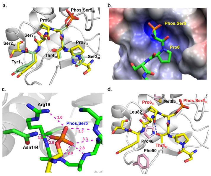
Figure 4.
Recognition of CTD by Drosophila melanogaster symplekin-Ssu72.
a. Superimposition of singly phosphorylated Ser5 CTD peptide bound at the active site of Drosophila melanogaster Ssu72 (peptide carbon shown in yellow) and human Ssu72 (peptide carbon shown in green). b. The electrostatic surface potential of active site of Drosophila Ssu72, where blue indicates positive charge, red indicates negative charge, and white indicates hydrophobic regions. The surfaces were generated with the Adaptive Poisson–Boltzmann Solver (APBS) using the AMBER force field (APBS Tools 2.1 PyMoL plugin, M. G. Lerner). c. A hydrogen bond network formed around phosphate group of phos.Ser5 CTD peptide in active site of Ssu72. d. Key residues of Drosophila Ssu72 for CTD recognition. The amino acids of Ssu72 are colored pink while the carbon atoms of CTD peptide are shown in yellow.

