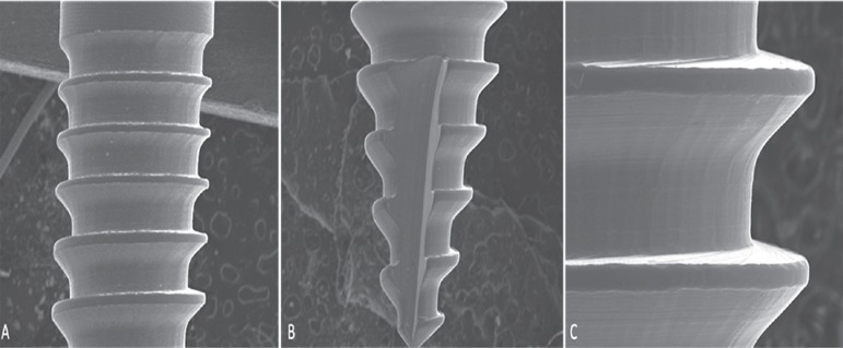
Figure 3.
Photomicrographs of the mini-implants studied. A) Area next to the transmucosal profile where mechanical fracture resistance test was performed (x50); B) Area next to the tip where mechanical fracture resistance test was performed (x50); C) Image of mini-implant body (x150). Smooth surface without defects, and tip with a cutting area.

