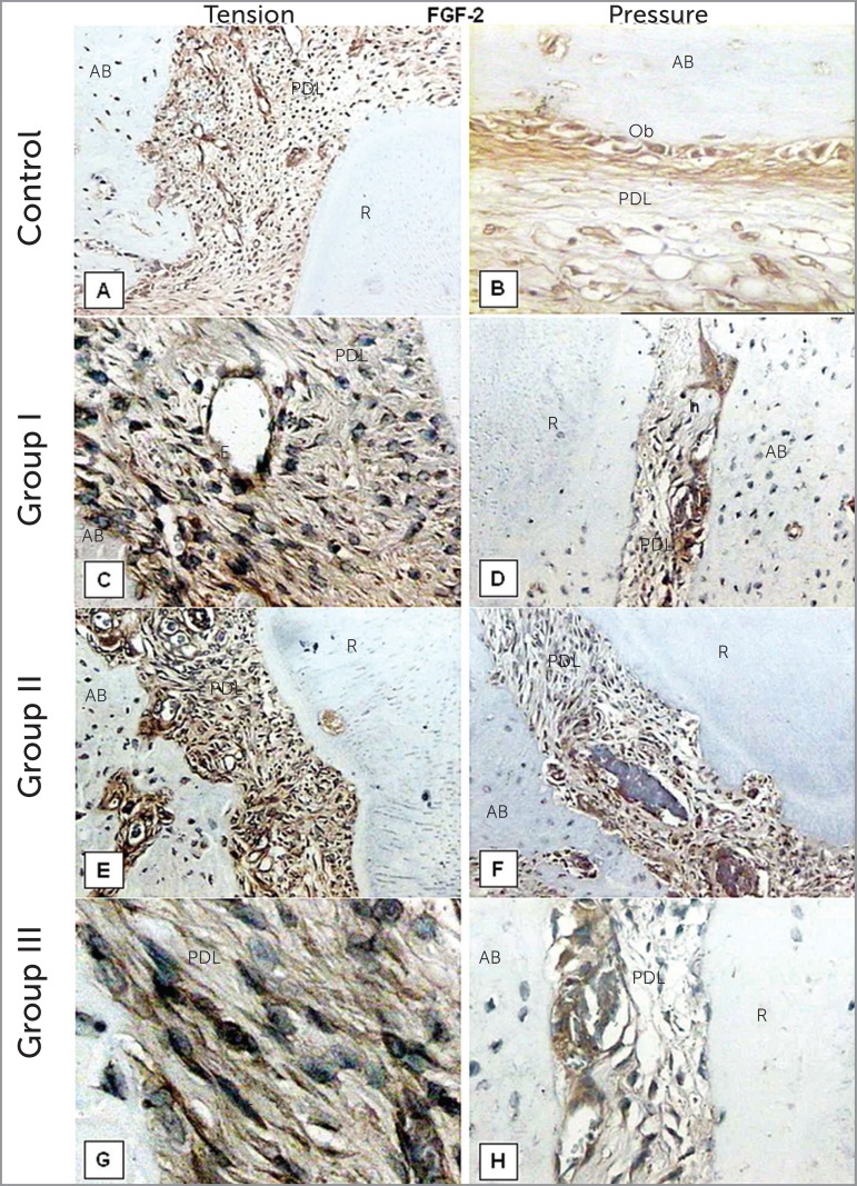
Figure 4.
FGF-2 immunohistochemistry staining of the control (A,B) and experimental groups (C-H) after 3, 7 and 14 days of orthodontic tooth movement. (A) magnification of 40x; (B, C, H) 200x; (D, E, F) 100x; (G) 400x. AB indicates alveolar bone; PDL, periodontal ligament; R, root; Ob, osteoblasts; E, endothelial cells; h, hialinized area; F, fibroblasts.

