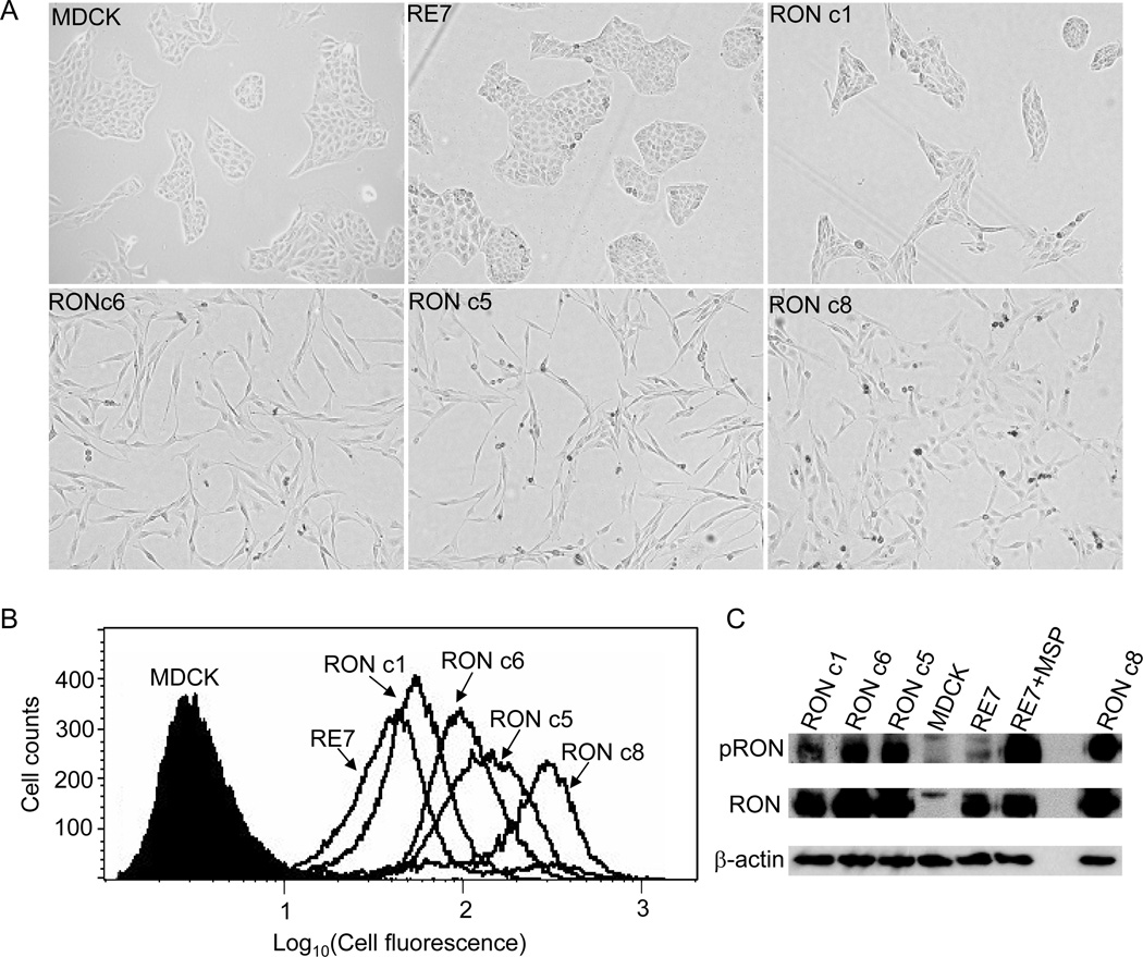
Fig. 1.
RON induces morphological changes and tyrosine phosphorylations in MDCK cells in proportion to its expression level. (A) Morphology of MDCK clones expressing RON. Pictures were taken 2 days after equal numbers of cells from each cell line were seeded. (B) RON expression on the cell surface was examined by FACS. (C) RON tyrosine phosphorylation was detected by 4G10 antibody (upper). The membrane was then stripped and reprobed with anti-RON C20 (lower).

