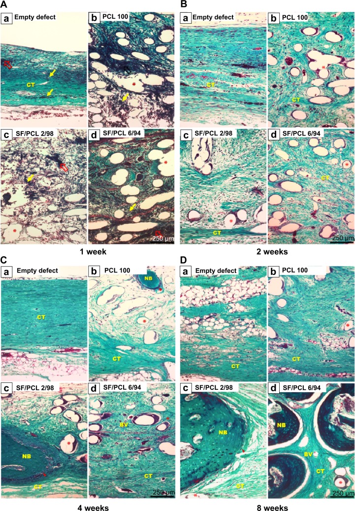
Figure 12.
Masson’s trichrome-stained histological section after implantation.
Notes: Representative histological sections show cross sections of the defect center after (A) 1 week, (B) 2 weeks, (C) 4 weeks, and (D) 8 weeks. Note the presence of blood clots (empty red arrows), inflammatory cells (thin yellow arrows), osteoblasts (arrowheads), and remaining scaffold (asterisks). Original magnification 100×.
Abbreviations: PCL, poly(ε-caprolactone); SF, silk fibroin; NB, new bone; BV, blood vessel; CT, connective tissue.

