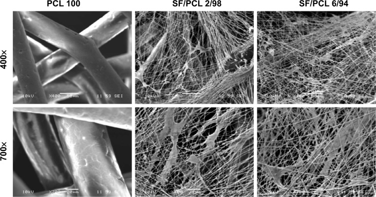
Figure 6.
Scanning electron micrographs of human mesenchymal stem cell-seeded scaffolds after 3 days of culturing.
Notes: Cell clusters were observed in the SF/PCL nano/microfibrous composite scaffolds, but not in the PCL microfibrous scaffold. Original magnification 400× and 700×.
Abbreviations: PCL, poly(ε-caprolactone); SF, silk fibroin.

