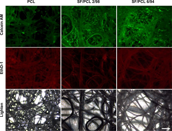
Figure 7.
Viability/cytotoxicity staining of cells cultured on PCL 100, SF/PCL 2/98, and SF/PCL 6/94 nano/microfibrous composite scaffolds were observed at 5 days of culturing.
Notes: Live cells were labeled with calcein acetomethoxy (AM; green), while dead cells were labeled with ethidium homodimer (EthD)-1 (red). Scale bar 200 μm.
Abbreviations: PCL, poly(ε-caprolactone); SF, silk fibroin.

