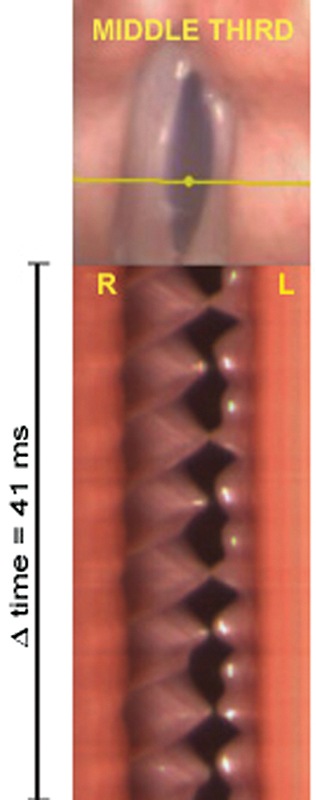
Fig. 10.

Digital videokymography obtained in the middle third of the glottis in case 4 (patient 4, with diagnosis of paralysis in of the left vocal fold). The free margin of the right (normal) vocal fold is spicule-shaped, and there is higher amplitude and irregular vibration in the left (paralyzed) vocal fold. Abbreviations: R, right; L, left; Δ time, time interval.
