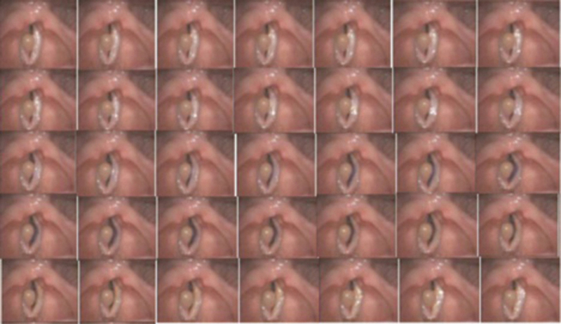
Fig. 7.

Successive frames obtained through digital videoendoscopy representing one complete glottal cycle (0.25 milliseconds intervals between frames) in case 3 (patient with an epidermoid cyst in the right vocal fold) and showing a difference in vibration amplitude between the right and left vocal folds.
