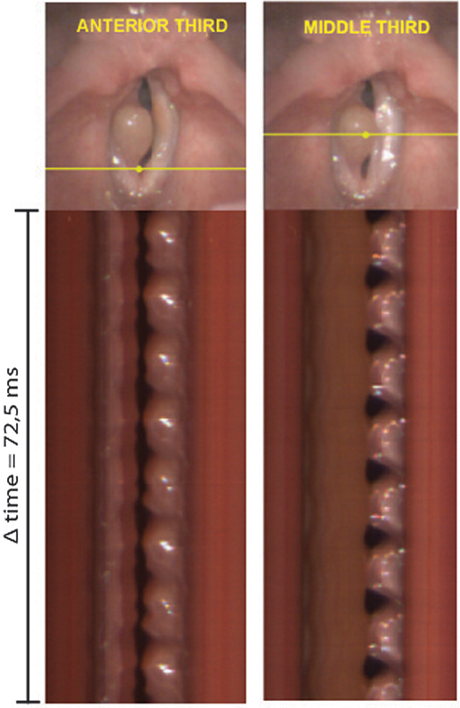
Fig. 8.

Digital videokymographic images of the anterior third (left) and middle third (anterior) of the vocal folds in case 3 (patient with an epidermoid cyst in the right vocal fold). In the middle third, the lesion prevents the mucosal wave. In the anterior middle, the bilobate outline of the left vocal fold indicates phase asymmetry within the same fold. Abbreviations: R, right; L, left; Δ time, time interval.
