
Fig. 1.
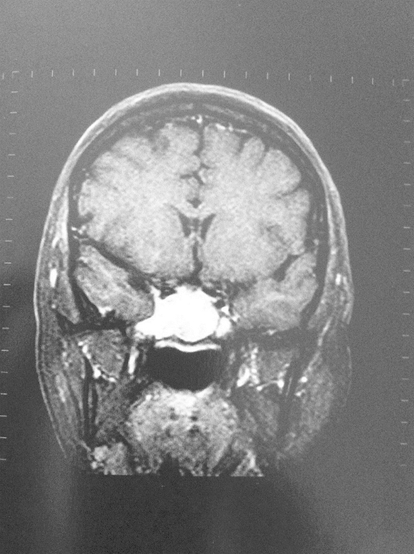
Coronal view of head magnetic resonance image reveals a lobulated mass occupying both sphenoid sinuses enhanced by the contrast media with no unequivocal evidence of skull base involvement.
Official websites use .gov
A
.gov website belongs to an official
government organization in the United States.
Secure .gov websites use HTTPS
A lock (
) or https:// means you've safely
connected to the .gov website. Share sensitive
information only on official, secure websites.
Coronal view of head magnetic resonance image reveals a lobulated mass occupying both sphenoid sinuses enhanced by the contrast media with no unequivocal evidence of skull base involvement.