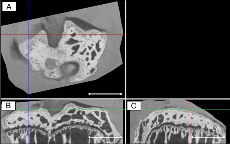
Figure 3.
A representative (A) cross-sectional, (B) coronal, and (C) sagittal contrast-enhanced nanofocus computed tomography (CE-nanoCT) image of a tibial plateau of a mouse of which 8 weeks in vivo medial meniscus destabilization (DMM) resulted in a local chondral defect (indicated by the crossed lines). Scale bars = 1 mm.

