Figure 3.
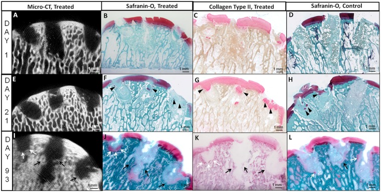
Micro-computed tomography images and corresponding Safranin O–stained and collagen type II–immunostained sections through chitosan-treated Jamshidi biopsy needle holes, and Safranin O images of matching contralateral controls in medial femoral condyles at day 1 (A-D), 3 weeks (E-H), and 3 months (I-L). Panels B to D and F to H were generated from cryosections and panels J to L from methylmethacrylate (MMA) sections. In treated condyles the left hole = 150 kDa chitosan, middle hole = 40 kDa chitosan, and right hole = 10 kDa chitosan. Evidence of bone remodeling and new bone growth (black arrows) as well as chondroinduction (white arrows) after 3 months is shown in panels I to L. Black arrowheads indicate articular cartilage fragments pushed into holes from adjacent cartilage.

