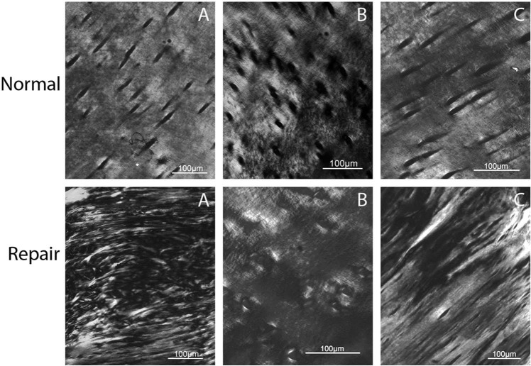
Figure 5.
Axial plane polarized light microscopy (PLM) images of the same 3 animals (A, B, C) and regions of repair and normal cartilage as presented in Figures 2-4. In all panels, superficial is toward the top of the image. PLM was able to detect a significant difference between repair and normal cartilage collagen structure. PLM repair scores: A= 0.5, B = 2.5, C = 2.5. PLM normal scores: A = 4, B = 4.5, C = 5.

