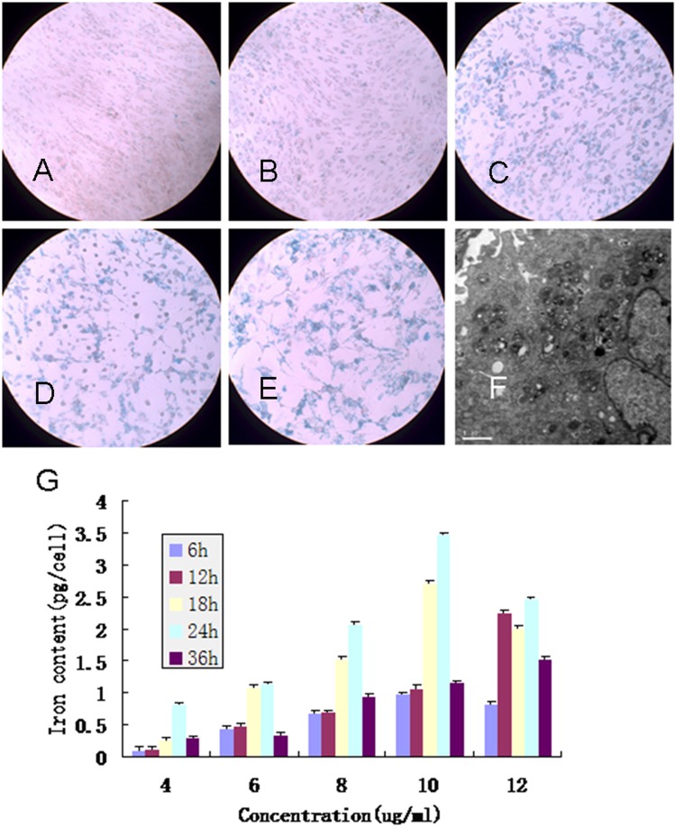
Figure 1.
(A-E) Prussian blue staining of green fluorescent protein (GFP) bone marrow mesenchymal stem cells (BMSCs) incubated with different concentrations of polyethylenimine-wrapped superparamagnetic iron oxide (PEI/SPIO) nanocomposites. (A) 4 µg/ml; (B) 6 µg/ml; (C) 8 µg/ml; (D) 10 µg/ml; (E) 12 µg/ml. (F) Transmission electron micrographs of GFP-BMSC labeled with 8 µg/ml SPIO (10,000x). (G) Cellular iron content of MSCs after labeling. The iron uptake process in MSCs displays a time- and dose-dependent behavior.

