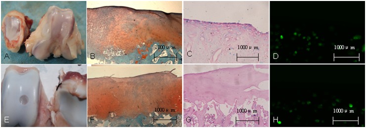
Figure 5.

Gross observation and histological evaluation of cell transplantation of superparamagnetic iron oxide (SPIO)-labeled bone marrow stem cells (BMSCs) in the knee of an experimental minipig model of articular cartilage defects at 24 weeks after surgery. (A, E) Gross observation for group A and group B. (B, F) Safranin-O staining showed proteoglycan deposition in trochlear groove defects at 24 weeks after surgery. (C, G) Prussian blue staining for the defect region. (D, H) Fluorescence microscopy demonstrated green fluorescence of BMSCs labeled with green fluorescent protein at defect region.
