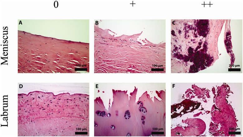
Figure 1.
Representative micrographs showing the degeneration of the menisci and labra graded 0, +, and ++. Degenerative changes were analyzed by light microscopy using sections stained with H&E. (A) Menisci with intact surfaces free of fibrillations were observed. (B) Degenerative surfaces with sharp fibrillations, continuous on one side and torn on the other side, exposing an undercut surface beneath the tear were also seen. (C) The most dramatic manifestation of degeneration was frank splitting of the matrix. Note the extensive calcification seen on this micrograph. (D) Labra with intact surfaces free of fibrillations were observed. (E) Degenerative labra exhibited acute, jagged surfaces. Note the chondrocytic proliferation and hyalinization seen on this micrograph. (F) Most dramatically, degenerative labra demonstrated frank tearing.

