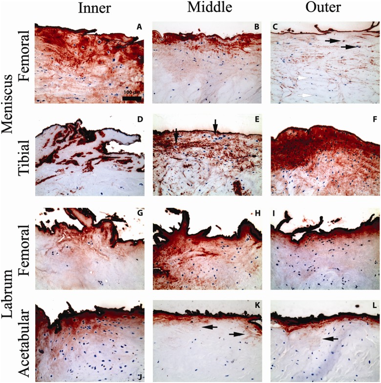
Figure 3.
Micrographs showing representative positive immunohistochemical staining for lubricin (red chromogen) at the surface of the tissues, in the extracellular matrix, and intracellularly in the menisci and labra. Micrographs are taken from the inner, middle, and outer thirds (segments) of the femoral and tibial sides of menisci and of the femoral and acetabular sides of labra. All micrographs are shown on the same scale. (A) Sample shows extensive matrix and intracellular staining for lubricin beneath a more pronounced surface-staining layer. (B) Matrix staining often follows the crimp pattern of collagen fibrils and fades with increasing depth into the tissue. (C) Lubricin staining is observed intracellularly in some cells (black arrows) but not in others (white arrows). (D) Lubricin staining is seen mainly as a surface coating in certain samples. (E) In some samples, lubricin in the matrix envelops the lacunae of chondrocyte-like cells, while the cell itself lacks intracellular staining (black arrows). (F) Lubricin is drastically seen here in a hypercellular region, fading out in deeper tissue. (G) Deposition of lubricin is seen in discrete granules within the extracellular matrix. (H) Lubricin is observed in granules in the matrix and intracellularly. (I) Granular deposition of lubricin in the matrix is most pronounced near the labral surface. (J) Intracellular lubricin is also more often observed near the labral surface. (K) Lubricin appears to diffuse into deeper tissue along the trabecular network of collagen fibers (black arrows). (L) Lubricin recapitulates the trabecular network of collagen fibers (black arrow).

