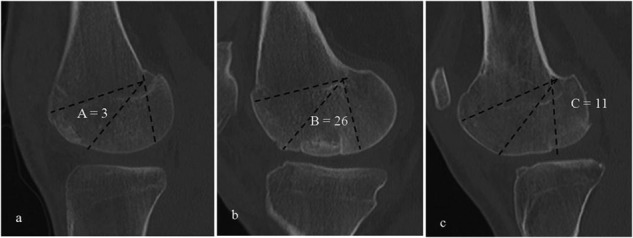
Figure 1.

Anatomic zones A (A), B (B), and C (C), as described by Cahill and Berg4 represented by computed tomography (CT) images. Zone B is considered the direct weightbearing zone, while zones A and C are regarded as indirect weightbearing areas. Each image contains one well-incorporated graft in the respective zone. Also shown is the number of grafts that were transplanted to each zone.
