
Figure 1.
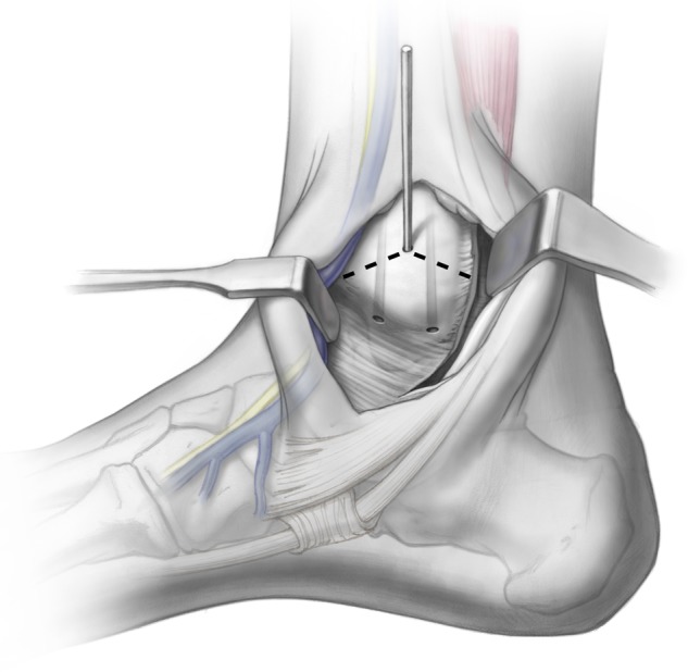
A provisional K-wire is drilled, exiting at the malleolar colliculus, to fluoroscopically visualize the medial malleolar osteotomy. A chevron-type cut is then made in the medial malleolus. Illustrations copyright of and reproduced with permission from JG Kennedy MD. Reproduction without express written consent is prohibited.