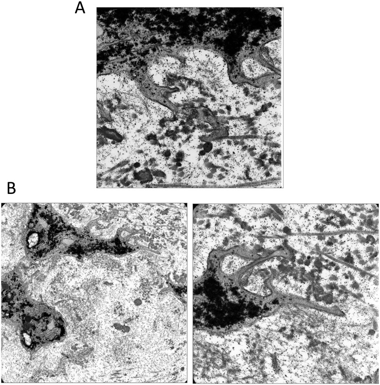
Figure 6.
Transmission electron micrographs showing chondrocyte processes adjacent to defects at 4 weeks after wounding. (A) A process has extended through the pericellular matrix and attached to collagen fibrils. (B) A different cell shows a larger process with smaller filipodia adhering directly to a collagen fibril.

