Figure 2.
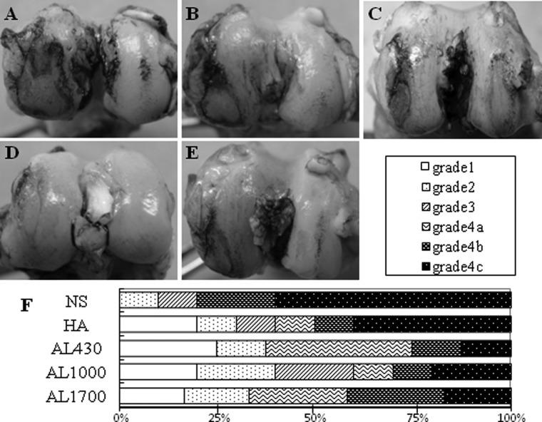
Macroscopic findings of femoral cartilage stained with India ink at 9 weeks after anterior cruciate ligament transection (ACLT) of (A) the normal saline (NS) group, (B) the hyaluronan (HA) group, (C) the AL430 group, (D) the AL1000 group, and (E) the AL1700 group. The retained intense black patches of ink indicate articular cartilage degeneration in the medial condyle. (F) Gross morphological grading of cartilage degeneration at the medial femoral condyle (MFC) in each group.

