
Figure 1.
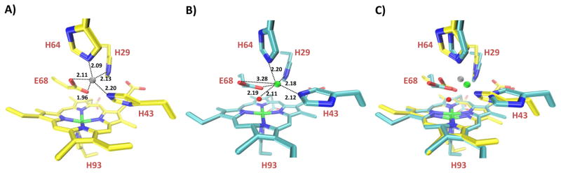
Crystal structure of rationally designed FeBMb overlays closely with minimized computer model. A) Minimized computer model of FeBMb with Zn(II) in the FeB site. B) Crystal structure of Fe(II)-FeBMb collected at Fe-edge absorption (1.7309 Å) at the Brookhaven National Synchrotron Light Source X12C beamline (Upton, NY) with 1.72 Å resolution. The Fe•••Fe distance is 4.82 Å, while the Fe-O-Fe angle is 115° (OE1 atom of E68). C) Overlay of FeBMb model (yellow) with Fe(II)-FeBMb crystal structure (cyan). In general, Fe(II) of the FeB site is represented by a green sphere; Zn(II) (grey sphere) was used to model the FeB site. A water molecule in the heme pocket is represented by a red sphere.