Figure 5.
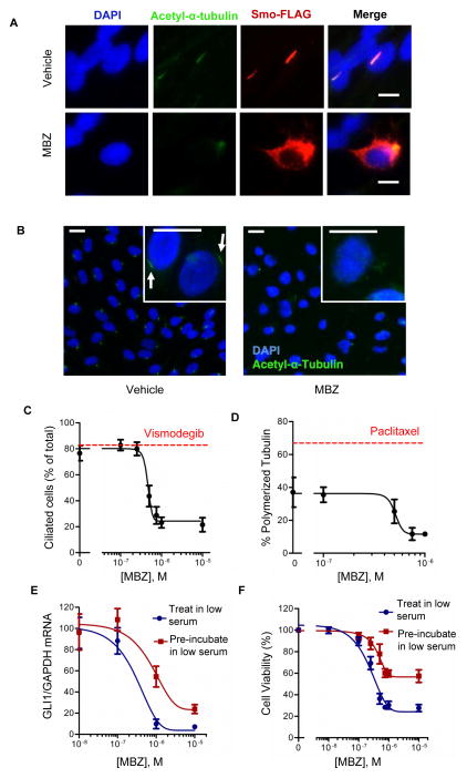
MBZ inhibits formation of primary cilia. (A) A Smo-FLAG fusion protein was expressed in hTERT-RPE1 cells by transient transfection. After 48 h incubation in low serum and treatment with 1 uM MBZ or vehicle, cells were fixed, permeabilized and stained with antibodies directed against acetyl-α-tubulin (green) and FLAG (red). Nuclei were counterstained with DAPI. Scale bar, 10 μm. (B) Cilia were numerically assessed by acetyl-α-tubulin staining in hTERT-RPE1 cells maintained in low serum conditions and treated with MBZ. Primary cilia (indicated by arrows) could be visualized on individual cells (inset). Scale bar, 20 μm. (C) The effects of MBZ or 0.2 μM vismodegib (red line) on the proportion of ciliated cells. (D) hTERT-RPE1 cells were treated with MBZ at the indicated concentrations or with 10 nM paclitaxel (red line) for 48 h under low serum conditions. Polymerized and unpolymerized tubulin fractions were quantified by immunobloting, normalized to the loading control β-actin, and expressed as the proportion of polymerized tubulin compared to the combined polymerized and unpolymerized tubulin. (E) GLI1 expression was assessed in DAOY cells that were incubated with MBZ for 48 h under the low serum conditions that allow formation of the primary cilium (“Treat in low serum”), or that were first maintained in low serum for 20 h prior to before adding MBZ for an additional 48 h (“Pre-incubate in low serum”). (F) Cell viability was assessed by CellTiter-Blue after the treatments described in (E).

