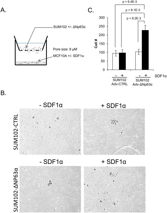
Figure 5. ΔNP63α positively regulates SDF1α dependent chemotaxis of breast cancer cells.
A) Diagram of the Boyden chamber assay. MCF10A cells (control and SDF1α overexpressing) were seeded in the bottom of the chamber and SUM102 cells (control or ΔNP63α overexpressing) were seeded onto the upper layer of the membrane. B) After 20 hours, the migrated cells were fixed and stained in crystal violet and where counted at 10 different fields. Graph represents the relative degree of migration based on the total number of cells counted. 10× magnification. C) Representative images of transmigrated cells per experimental group. Experiments were performed in triplicate (n=3), data are means ± S.D.; p-values are indicated.

