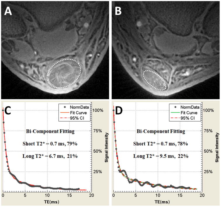
Figure 10.

60-year old man several years after successful right Achilles tendon repair. Axial UTE MR images of the repaired right Achilles tendon (A) compared with the same patient's asymptomatic left Achilles tendon (B). Quantitative bi-component analysis performed in the tendons (dashed ovals) show that the short T2* value and fraction of the repaired right tendon (C) has approached the asymptomatic left side (D), confirming adequate collagen remodeling.
