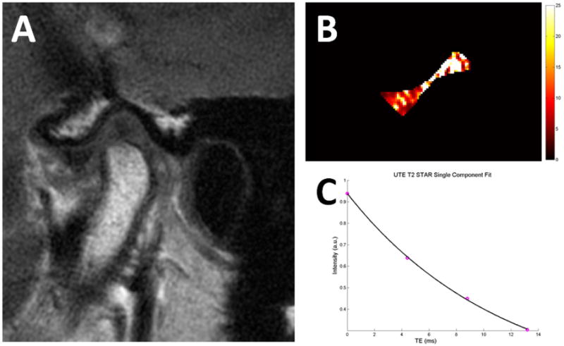
Figure 9.

Temporomandibular joint disc of a 35-year old asymptomatic volunteer. (A) Conventional T1-weighted fast-spin echo image with mouth closed (A) shows the normal position of the TMJ disc. Quantitative pixel map generated from UTE images (B) show increased T2* values at the intermediate zone and posterior band, suggestive of early-stage degeneration. Mono-exponential T2* analysis of the entire disc (C) shows excellent curve fitting with T2* value of 11.4 ms.
