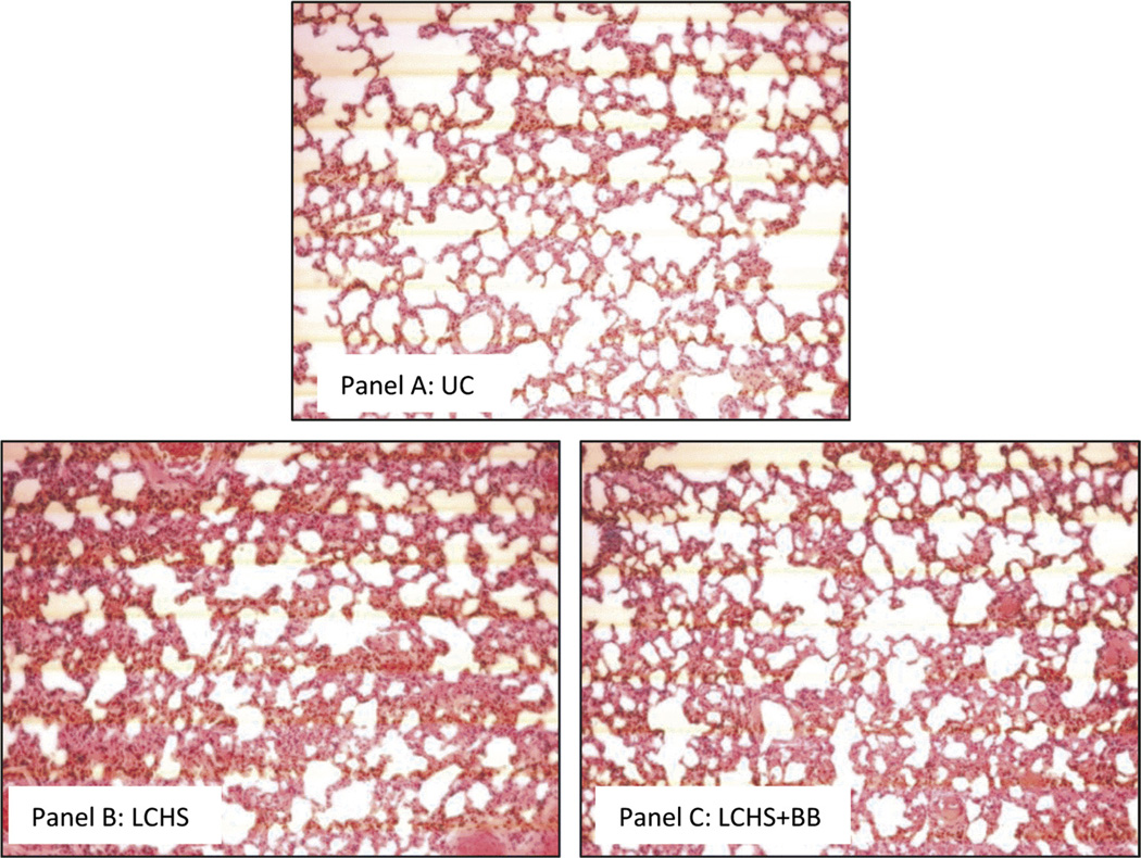
Figure 3.
A–C, Effect of propranolol on histologic lung injury after trauma and shock. Sections of contused lung were stained with hematoxylin and eosin. Images are taken at 20× magnification. A, Representative image from a naive control animal with normal lung architecture. B, Representative image from an animal after LCHS showing hemorrhage, increased inflammatory cells, edema, and disruption of alveoli. C, Representative image from animal receiving BB after LCHS and demonstrates hemorrhage, with less edema and less inflammatory cells. n = 6 animals per group.

