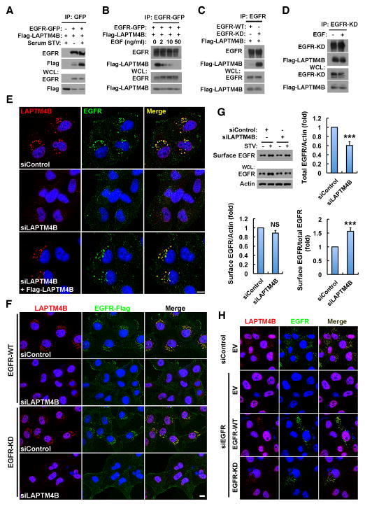
Figure 3. Inactive EGFR and LAPTM4B Interact and Stabilize Each Other at Endosomes.
(A) Co-immunoprecipitation (co-IP) of EGFR-GFP with Flag-LAPTM4B in HEK 293 cells cultured in normal or serum free medium.
(B) Co-IP of EGFR-GFP with Flag-LAPTM4B in HEK 293 cells cultured in serum free medium with 0, 2, 10, or 50 ng/ml EGF treatment (30 min).
(C) Co-IP of wild type (WT) or kinase dead (KD) EGFR with Flag-LAPTM4B in HEK 293 cells in normal medium.
(D) Co-IP of EGFR-KD with Flag-LAPTM4B in HEK 293 cells cultured in serum free medium with or without 50 ng/ml EGF treatment (30 min).
(E) LAPTM4B is required for the endosomal accumulation of endogenous EGFR. MDA-MB-231 cells were treated with control or LAPTM4B siRNA, and after 48 h the siRNA-resistant LAPTM4B was re-expressed via transient transfection. Cells were serum starved and fixed for co-staining of LAPTM4B (red) and EGFR (green).
(F) Ectopically expressed wild type (WT) and kinase dead (KD) EGFR depend on LAPTM4B for endosomal accumulation. MDA-MB-231 cells stably expressing C-terminally Flag-tagged EGFR-WT or -KD were treated with control or LAPTM4B siRNA, serum starved and fixed for co-staining of Flag (green) and endogenous LAPTM4B (red).
(G) Control and LAPTM4B knockdown cells were surface biotinylated, and surface and total EGFR levels were analyzed by western blot (top left). Quantification of total EGFR levels (top right), cell surface EGFR levels (bottom left), and the relative amounts of cell surface EGFR (bottom right) in serum starved control and LAPTM4B knockdown cells; mean + SD, n = 3; *** P < 0.001; NS, not significant.
(H) Inactive EGFR stabilizes endosomal LAPTM4B. MDA-MB-231 cells were transfected with control or EGFR siRNA, and after 48 h cells were transfected with empty vector (EV) or siRNA-resistant EGFR-WT or -KD and starved overnight, followed by fixing and co-staining of EGFR (green) and LAPTM4B (red).
WCL, whole cell lysate; STV, serum starvation; Bar, 10 μm; DAPI was used to stain the nuclei. See also Figure S3.

