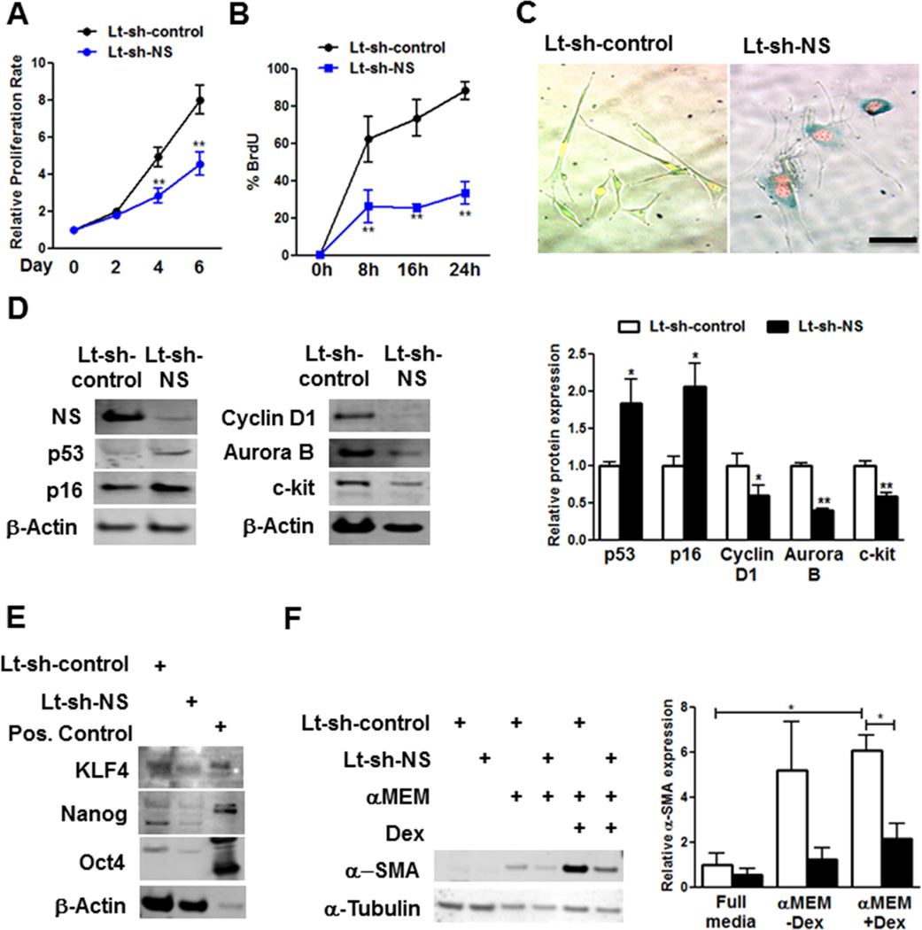
FIGURE 3. NS Silencing Causes Cell Cycle Arrest and Senescence in YCPCs.
(A) Proliferation rate determined by Cyquant assay (N = 5) (B) BrdU incorporation measured by fluorescence activated cell sorting (FACS) (N = 3). (C) Images of YCPCs expressing fluorescence ubiquitination cell cycle indicator constructs stained with SA-β-gal. (D) Immunoblot and densitmetric analyses of p53 (N = 13), p16 (N = 5), Cyclin D1 (N = 3), Aurora B (N = 3), and c-kit (N = 6) in YCPCs. (E) Immunoblots of multipotency markers KLF4, Nanog, and Oct 4 in YCPCs. Positive (Pos.) control: mouse embryonic stem cell lysate. (F) Immunoblot and densitometric analyses of α-smooth muscle actin (α-SMA) expression in YCPCs cultured in normal or minimal essential media (αMEM) with or without dexamethasone (Dex) (N = 3). Scale bar: 100γm.
*p < 0.05; **p < 0.01

