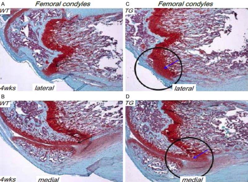
Figure 3.

Less cartilage resorption in Col10a1-Runx2 mice. (A, B) Safranin O/fast green staining of sagittal section of femoral condyle from 4 wks WT mouse show clear cartilage resorption at the frontal margins of the proximal lateral (A) and medial (B) femoral condyles (black arrows). (C, D) Compared to the WT littermate controls, TG mice showed a much decreased cartilage resorption in corresponding lateral and medial femoral condyles (blue arrows).
