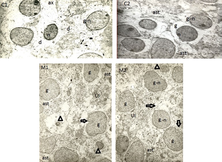
Fig 1.
Representative electron microscope (EM) photomicrographs of fetal parietal cortex in rats. The images C1 and C2 belong to two fetuses from control rats, and the images M1 and M2 belong to a music-treated group. Shape of the cells was simpler and smoother in control rats, whereas the cell and nuclear membranes are more complex in the music-treated group. " g" indicates granular cell, " n" stands for nucleus, " ast" indicates astrocyte, " UI" stands for unidentified cells,⇨ indicates indentation in cell membrane and/or nucleus membrane, △ indicates nucleus of unknown cells, " d" stands for dendrite, and " ax" indicates axon.

