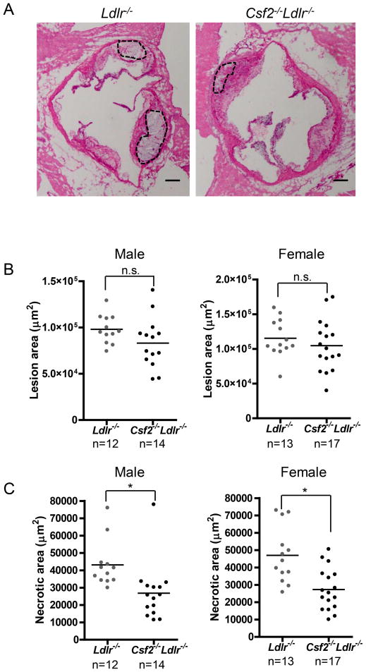
Figure 1. Necrotic area is decreased in the aortic root lesions of GM-CSF-deficient Ldlr−/− mice.
A, Representative images of H&E-stained aortic root sections of 12-wk WD-fed Ldlr−/− and Csf2−/−Ldlr−/− mice. The necrotic regions are indicated by the broken lines. Bar, 50 μm. B–C, Measurement of total atherosclerotic lesion area and necrotic area. *, p<0.05; n.s., no significant difference.

