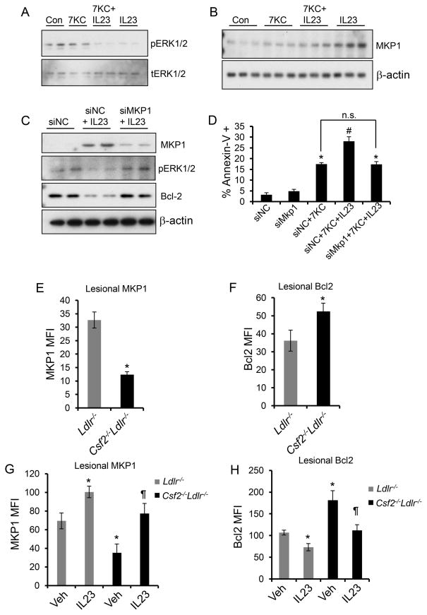
Figure 7. Evidence that an increase in MKP-1 links GM-CSF/IL-23 to decreased Bcl-2 and apoptosis in macrophages and in atherosclerotic lesions.
A, Macrophages treated with 7KC alone, IL-23 alone, or the combination of 7KC and IL-23 for 30 min were probed for phospho- or total ERK1/2 by immunoblotting. B, Immunoblot of MKP-1 in macrophages treated with 7KC alone, IL-23 alone, or the combination of 7KC and IL-23 for 4 h. C, Macrophages were transfected with negative control siRNA (siNC) or siRNA against MKP-1 (siMkp1) and then treated 48 h later without or with IL-23 for 8 h. Whole cell lysates were immunoblotted for MKP-1, pERK1/2, Bcl-2, and β-actin. D, Macrophages transfected with siNC or siMkp1 were treated with 7KC or a combination of 7KC and IL-23 for 18 h followed by quantification of apoptosis by microscopic analysis of annexin V-labeled macrophages. E-H, Immunofluorescence quantification (mean fluorescence intensity, MFI) of MKP-1 and Bcl-2 in F4/80+ macrophage-rich regions of aortic root atherosclerotic lesions. n=6 mice per group. In G and H, the indicated groups of mice were treated with saline (Veh) or rIL-23 (5 μg/kg). In D, the data are representative of 3 independent experiments. In E and F, n=6 mice per group, and in G and H, n=3 mice for the control group and n=6 mice for rIL-23-treated group; *, p<0.05 vs. control or Ldlr−/− mice; #, p<0.05 vs. 7KC treatment; ¶, p<0.05 vs. vehicle-treated Csf2−/−Ldlr−/− mice; n.s., no significant difference.

