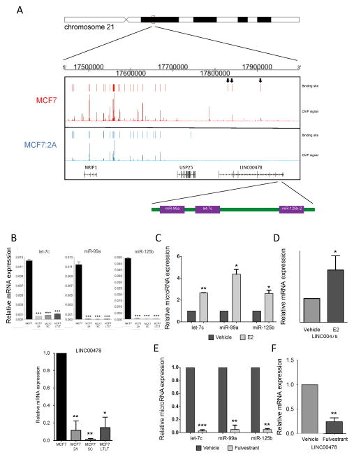
Figure 2. The let-7c/miR-99a/miR-125b cluster is regulated by the ER.
A) The top panel represents a schematic of the genomic location of the let-7c/miR-99a/miR-125b cluster within chromosome 21. The ER ChIP-Seq signal derived from both MCF7 (shown in red) and MCF7:2A (shown in blue) cells is shown demonstrating a loss of ER signal at the loci near the let-7c/miR-99a/miR-125b cluster. The ER binding sites within LINC00478 lost in MCF7:2A cells are indicated with arrows. B) The relative expression level of let-7c, miR-99a, and miR-125b (top) and LINC00478 (bottom) is shown in the MCF7, MCF7:2A, MCF7:5C, and MCF7:LTLT cell lines. C) and D) E2 regulates the expression of the cluster miRNAs and primary transcript. MCF7 cells were treated with E2 for 3 h, and the level of let-7c, miR-99a, miR-125b, and LINC00478 expression was determined by RT-PCR. E) and F) Fulvestrant treatment leads to loss of the cluster miRNAs and LINC00478. MCF7 cells were treated with fulvestrant for 48 h, and the level of let-7c, miR-99a, miR-125b, and LINC00478 expression was determined by RT-PCR. *, p < 0.01; **, p < 0.01, ***; p < 0.0001.

