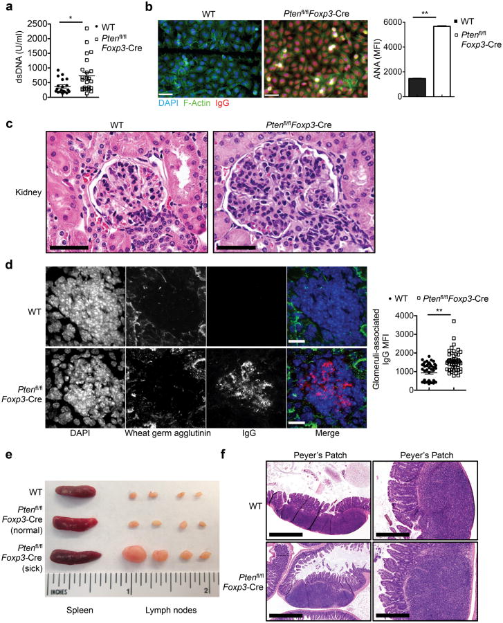
Figure 1. Ptenfl/flFoxp3-Cremice develop age-related autoimmune and lymphoproliferative disease.
(a) Quantification of dsDNA-specific IgG in the serum of Pten+/+Foxp3-Cre (WT) and Ptenfl/flFoxp3-Cre mice (2-6 months old). (b) Representative images (scale 60 μm) and quantification of fluorescent intensity (right) of serum ANA IgG autoantibodies detected with fixed Hep-2 slides. (c) Histology images of kidney glomeruli sections stained with H&E (magnification: ×60; scale 50 μm). (d) Immune fluorescence images of kidney glomeruli sections showing IgG deposits (scale 40 μm). (e) Images of spleen and peripheral lymph nodes from WT (upper, ∼5 months old), Ptenfl/flFoxp3-Cre mice prior to the development of lymphoproliferative disease (middle, ∼2.5 months old), and Ptenfl/flFoxp3-Cre mice with lymphoproliferative disease (lower, ∼5 months old). (f) H&E staining of Peyer's patches in the intestine of WT and Ptenfl/flFoxp3-Cre mice (magnification: left, ×4; scale 1mm and right, ×20; scale 200 μm). Data are representative of at least two independent experiments (a-f). *P < 0.05 and **P < 0.001. Data are mean ± s.e.m.

