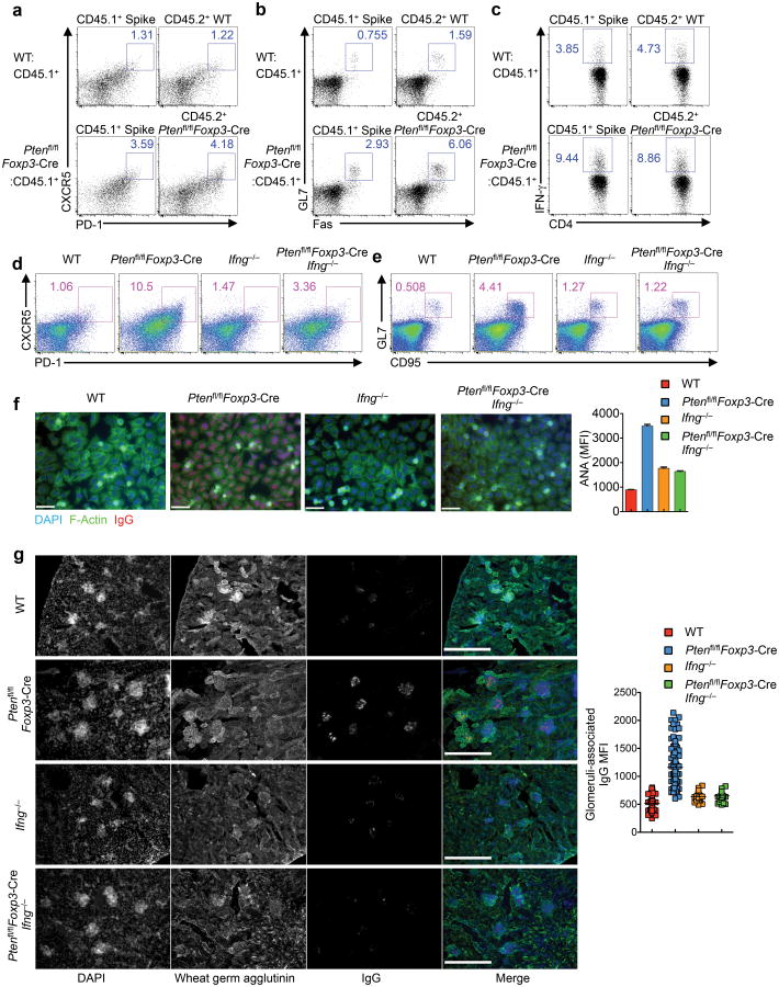
Figure 4. Analysis of bone marrow-derived chimeras and Ptenfl/flFoxp3-Cre Ifng−/− mice reveals an important contribution of IFN-γ overproduction to dysregulated TFH responses in Ptenfl/flFoxp3-Cre mice.
(a-c) Sublethally irradiated Rag1−/− mice were reconstituted with a 1:1 mix of CD45.1+ BM and either CD45.2+ WT or Ptenfl/flFoxp3-Cre BM cells. Following reconstitution, the mixed chimeras were analyzed for TFH (a), GC B cells (b), and intracellular staining of IFN-γ in CD4+ T cells (c). (d,e) Analysis of TFH (d) and GC B cells (e) in the spleen of WT, Ptenfl/flFoxp3-Cre, Ifng−/− and Ptenfl/flFoxp3-Cre Ifng−/− mice. (f) Representative images and quantification of fluorescent intensity (right) of ANA IgG autoantibodies detected with Hep-2 slides in the serum from WT, Ptenfl/flFoxp3-Cre, Ifng−/− and Ptenfl/flFoxp3-Cre Ifng−/− mice (scale 60 μm). (g) Representative images of immune fluorescence imaging of kidney sections showing IgG deposits (scale 300 μm), and quantitative analysis (right). Data are representative of three independent experiments (a-e) and one experiment (f,g; n=3 WT, 6 Ptenfl/flFoxp3-Cre, 2 Ifng−/− and 3 Ptenfl/flFoxp3-Cre Ifng−/− mice). Data are mean ± s.e.m.

