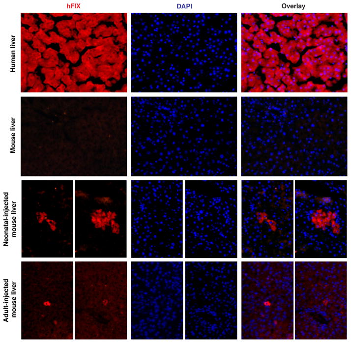
Extended Data Figure 1. hF9 liver immunohistochemistry.
From top to bottom, panels show human factor IX staining (red) with DAPI nuclear counterstain (blue) in positive control human liver, negative control untreated mouse liver, and two sets of representative stains from mice treated as neonates or adults with AAV8-P2A-hF9.

