
Fig 4. Shifting fates: cartilage to bone.
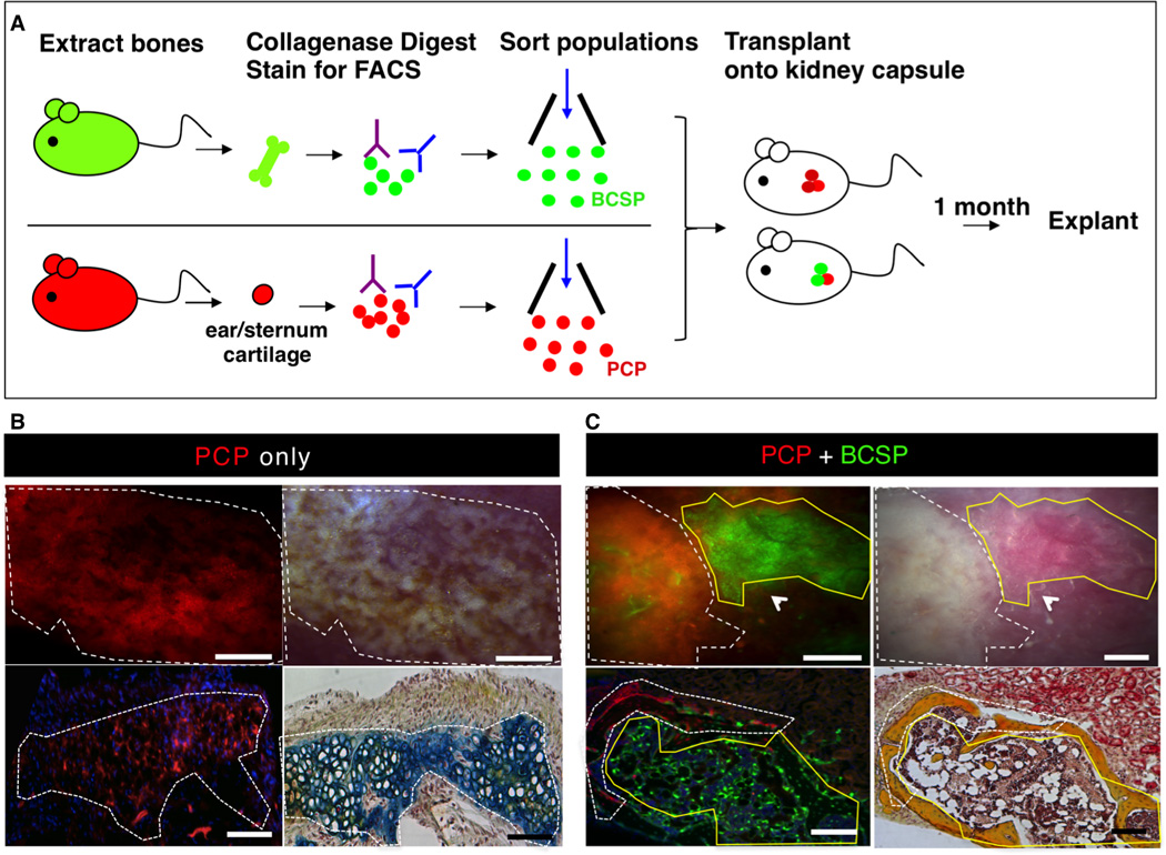
(A) Scheme of experiment: BCSPs were isolated from femora of GFP+ mice at P3 and PCPs (pro-chondrogenic progenitor) were isolated from ears/sternum of RFP+ adult mice following mechanical and enzymatic digestion, and subsequent FACS fractionation. Purified GFP+ BCSP and RFP+ PCP were co-transplanted beneath the kidney capsules of immunodeficient recipient mice. RFP+ PCP transplant served as a control. 1 month after transplantation, all grafts were removed for analysis.
(B) Microscopy of explanted grafts 1 month following transplantation of 20,000 RFP+ PCP cells (see Figure 4A). The white dotted line outlines the extent of the graft formed. Fluorescence micrograph of explanted graft demonstrating the presence of an RFP+ graft (upper left). Corresponding brightfield micrograph is shown in upper right panel. Transverse section stained with Movat’s Pentachrome demonstrates that RFP-labeled PCP cells form cartilage (blue stain) (lower right). Scale bar: 500µm (upper panel), 200µm (lower panel). Representative of 4 replicates.
(C) Microscopy of explanted grafts following co-transplantation of 20,000 RFP-labeled PCP cells with 20,000 GFP-labeled BCSPs (as detailed in Figure 4A). The grafts, which formed, are outlined by a white dotted line (indicating RFP+ portion) and a yellow solid line (indicating GFP+ portion) (upper left). A corresponding brightfield micrograph is shown in the upper right panel. A transverse section stained with Movat’s pentachrome shows that both RFP-labeled PCPs and GFP-labeled BCSPs form bone (yellow stain) (lower right). Scale bar: 500µm (upper panel), 200µm (lower panel). Representative of 5 replicates.