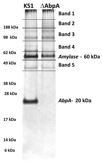
Figure 3. Sample preparation for MS/MS analysis of amylase-precipitated proteins.
Coomassie-stained gels were cut in the pattern depicted above to get 5 bands/slices per strain. Amylase and AbpA bands were avoided as they are well known from past experiments. The gel bands/slices were placed in microcentrifuge tubes containing 70 μl of double distilled water and sent for nano-LC/MS/MS analysis.

