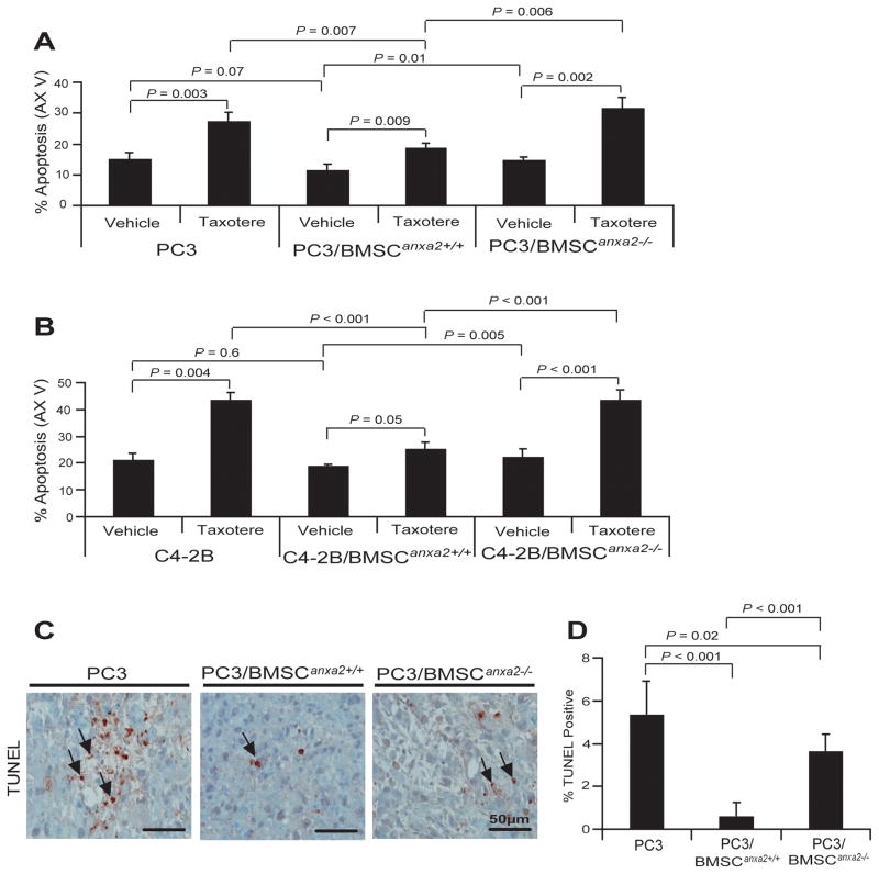
Figure 4. ANXA2 and CXCL12 in the marrow environment enhance PCa survival and drug-resistance.
PC3GFP or C4-2BGFP cells were directly cultured with BMSCAnxa2+/+ or BMSCAnxa2−/− for 48 hours and taxotere was introduced into the culture for an additional 48 hours. Annexin V staining was performed on the recovered (A) PC3 or (B) C4-2B cells and quantified by FACS. Data in (Fig. 4A and B) are representative of mean with s.d. for triplicates in each of three independent experiments (Student’s t-test). (C) TUNEL staining of apoptotic PCa cells (black arrows) of tumors grown from PC3luc cells alone or mixed with BMSCAnxa2+/+ or BMSCAnxa2−/− implanted into SCID mice. BAR = 50μm. (D) Quantification of % TUNEL positive cells in Fig. 4C (mean±s.d., Student’s t-test).

