
Fig. 1.
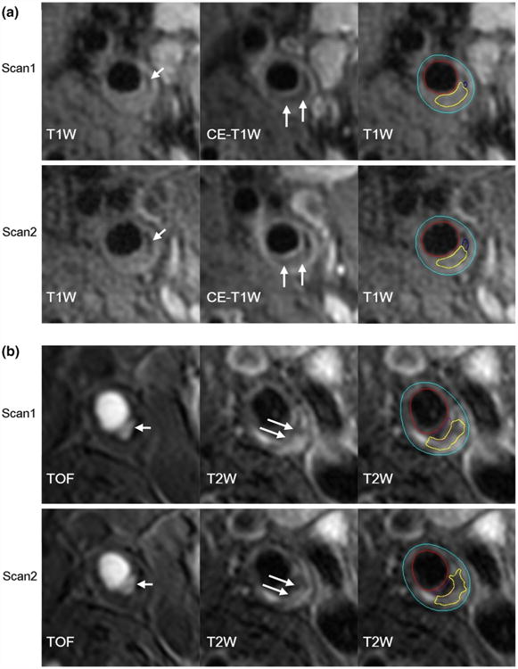
First and repeat scans on different scanner platforms. a A plaque with calcification (short arrows) and LRNC (long arrows) was scanned twice on GE platform. b A plaque with ulceration (short arrows) and LRNC (long arrows) was scanned twice on Philips platform. Primary weightings are presented to highlight specific features. The last image in each panel illustrates contour-based measurements (yellow contours indicate LRNC). CE-T1W contrast-enhanced T1-weighted, LRNC lipid-rich necrotic core, T1W T1-weighted, T2W T2-weighted, TOF time-of-flight